Explore
 Validate
 Learn
Learn Western blot
 Immunohistochemistry
Antibody data
- Antibody Data
- Antigen structure
- References [0]
- Comments [0]
- Validations
- Western blot [2]
- Immunocytochemistry [3]
- Immunohistochemistry [11]
Submit
Validation data
Reference
Comment
Report error
- Product number
- LS-C312942 - Provider product page

- Provider
- LSBio
- Product name
- SSR3 / TRAP-Gamma Antibody (aa8-23) LS-C312942
- Antibody type
- Polyclonal
- Description
- Immunogen affinity purified
- Reactivity
- Human, Mouse, Rat
- Host
- Rabbit
- Storage
- At -20°C for 1 year. After reconstitution, at 4°C for 1 month. It can also be aliquotted and stored frozen at -20°C for a longer time. Avoid freeze-thaw cycles.
No comments: Submit comment
Supportive validation
- Submitted by
- LSBio (provider)
- Enhanced method
- Genetic validation
- Main image
- Experimental details
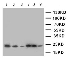
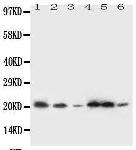
- WB of SSR3 / TRAP-Gamma antibody. Lane 1: Rat Liver Tissue Lysate. Lane 2: Rat Spleen Tissue Lysate. Lane 3: A431 Cell Lysate. Lane 4: HELA Cell Lysate. Lane 5: U87 Cell Lysate. Lane 6: SMMC Cell Lysate.
- Submitted by
- LSBio (provider)
- Enhanced method
- Genetic validation
- Main image
- Experimental details
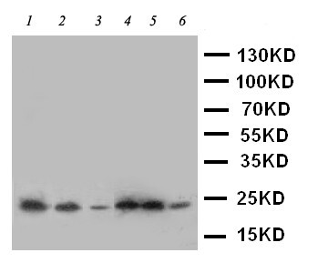
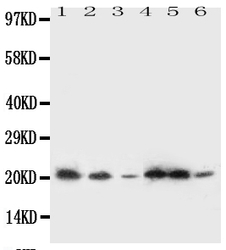
- Anti-SSR3 antibody, Western blotting All lanes: Anti SSR3 at 0.5ug/ml Lane 1: Rat Liver Tissue Lysate at 50ugLane 2: Rat Spleen Tissue Lysate at 50ugLane 3: A431 Whole Cell Lysate at 40ugLane 4: HELA Whole Cell Lysate at 40ugLane 5: U87 Whole Cell Lysate at 40ugLane 6: SMMC Whole Cell Lysate at 40ugPredicted bind size: 21KD Observed bind size: 21KD
Supportive validation
- Submitted by
- LSBio (provider)
- Enhanced method
- Genetic validation
- Main image

- Experimental details
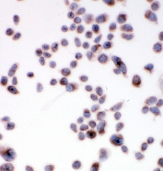
- ICC analysis of SSR3 using anti-SSR3 antibody. SSR3 was detected in immunocytochemical section of HELA cell. Heat mediated antigen retrieval was performed in citrate buffer (pH6, epitope retrieval solution) for 20 mins. The tissue section was blocked with 10% goat serum. The tissue section was then incubated with 1µg/ml rabbit anti-SSR3 Antibody overnight at 4°C. Biotinylated goat anti-rabbit IgG was used as secondary antibody and incubated for 30 minutes at 37°C. The tissue section was developed using Strepavidin-Biotin-Complex (SABC) with DAB as the chromogen.

- Submitted by
- LSBio (provider)
- Main image
- Experimental details
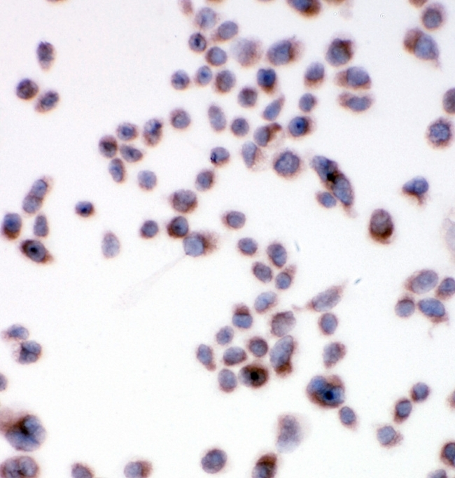
- ICC analysis of SSR3 using anti-SSR3 antibody. SSR3 was detected in immunocytochemical section of HELA cell. Heat mediated antigen retrieval was performed in citrate buffer (pH6, epitope retrieval solution) for 20 mins. The tissue section was blocked with 10% goat serum. The tissue section was then incubated with 1µg/ml rabbit anti-SSR3 Antibody overnight at 4°C. Biotinylated goat anti-rabbit IgG was used as secondary antibody and incubated for 30 minutes at 37°C. The tissue section was developed using Strepavidin-Biotin-Complex (SABC) with DAB as the chromogen.
- Submitted by
- LSBio (provider)
- Main image

- Experimental details
- ICC analysis of SSR3 using anti-SSR3 antibody. SSR3 was detected in immunocytochemical section of HELA cell. Heat mediated antigen retrieval was performed in citrate buffer (pH6, epitope retrieval solution) for 20 mins. The tissue section was blocked with 10% goat serum. The tissue section was then incubated with 1µg/ml rabbit anti-SSR3 Antibody overnight at 4°C. Biotinylated goat anti-rabbit IgG was used as secondary antibody and incubated for 30 minutes at 37°C. The tissue section was developed using Strepavidin-Biotin-Complex (SABC) with DAB as the chromogen.

Enhanced validation
- Submitted by
- LSBio (provider)
- Enhanced method
- Genetic validation
- Main image
- Experimental details
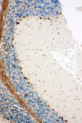
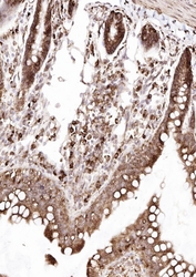
- SSR3 / TRAP-Gamma antibody. IHC(P): Rat Brain Tissue.

- Submitted by
- LSBio (provider)
- Enhanced method
- Genetic validation
- Main image

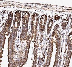
- Experimental details
- SSR3 / TRAP-Gamma antibody. IHC(P): Rat Intestine Tissue.

- Submitted by
- LSBio (provider)
- Enhanced method
- Genetic validation
- Main image
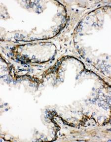
- Experimental details
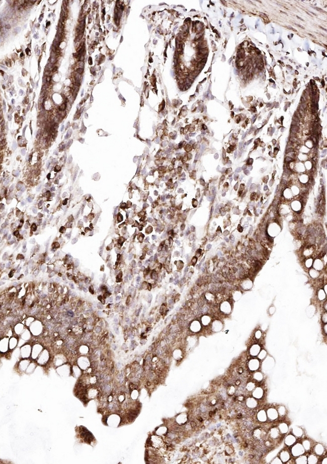
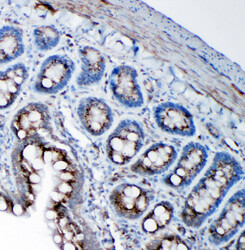
- IHC analysis of SSR3 using anti-SSR3 antibody. SSR3 was detected in paraffin-embedded section of mouse intestine tissues. Heat mediated antigen retrieval was performed in citrate buffer (pH6, epitope retrieval solution) for 20 mins. The tissue section was blocked with 10% goat serum. The tissue section was then incubated with 1µg/ml rabbit anti-SSR3 Antibody overnight at 4°C. Biotinylated goat anti-rabbit IgG was used as secondary antibody and incubated for 30 minutes at 37°C. The tissue section was developed using Strepavidin-Biotin-Complex (SABC) with DAB as the chromogen.

- Submitted by
- LSBio (provider)
- Enhanced method
- Genetic validation
- Main image

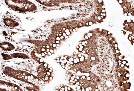
- Experimental details
- IHC analysis of SSR3 using anti-SSR3 antibody. SSR3 was detected in paraffin-embedded section of rat intestine tissues. Heat mediated antigen retrieval was performed in citrate buffer (pH6, epitope retrieval solution) for 20 mins. The tissue section was blocked with 10% goat serum. The tissue section was then incubated with 1µg/ml rabbit anti-SSR3 Antibody overnight at 4°C. Biotinylated goat anti-rabbit IgG was used as secondary antibody and incubated for 30 minutes at 37°C. The tissue section was developed using Strepavidin-Biotin-Complex (SABC) with DAB as the chromogen.

- Submitted by
- LSBio (provider)
- Enhanced method
- Genetic validation
- Main image

- Experimental details
- IHC analysis of SSR3 using anti-SSR3 antibody. SSR3 was detected in paraffin-embedded section of rat intestine tissues. Heat mediated antigen retrieval was performed in citrate buffer (pH6, epitope retrieval solution) for 20 mins. The tissue section was blocked with 10% goat serum. The tissue section was then incubated with 1µg/ml rabbit anti-SSR3 Antibody overnight at 4°C. Biotinylated goat anti-rabbit IgG was used as secondary antibody and incubated for 30 minutes at 37°C. The tissue section was developed using Strepavidin-Biotin-Complex (SABC) with DAB as the chromogen.
- Submitted by
- LSBio (provider)
- Enhanced method
- Genetic validation
- Main image

- Experimental details
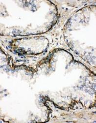
- SSR3 / TRAP-Gamma antibody. IHC(P): Human Prostatic Cancer Tissue.
- Submitted by
- LSBio (provider)
- Enhanced method
- Genetic validation
- Main image

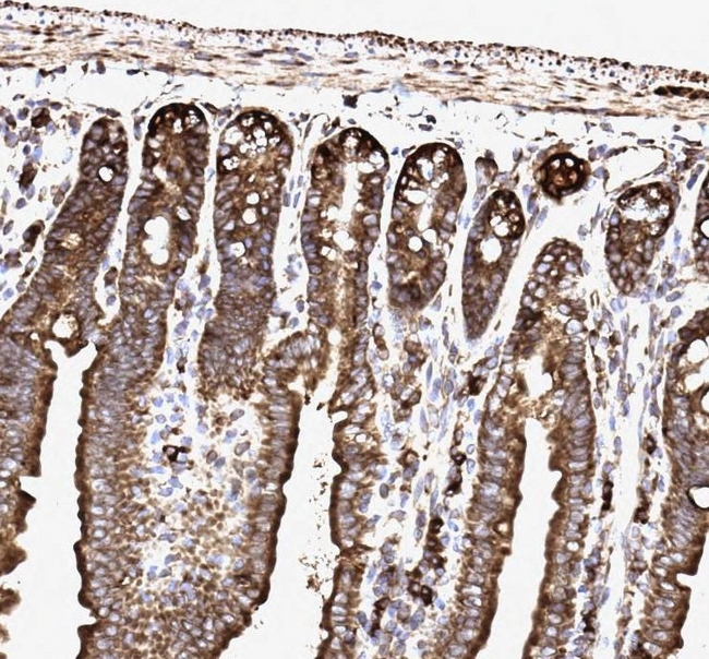
- Experimental details
- IHC analysis of SSR3 using anti-SSR3 antibody. SSR3 was detected in paraffin-embedded section of mouse intestine tissues. Heat mediated antigen retrieval was performed in citrate buffer (pH6, epitope retrieval solution) for 20 mins. The tissue section was blocked with 10% goat serum. The tissue section was then incubated with 1µg/ml rabbit anti-SSR3 Antibody overnight at 4°C. Biotinylated goat anti-rabbit IgG was used as secondary antibody and incubated for 30 minutes at 37°C. The tissue section was developed using Strepavidin-Biotin-Complex (SABC) with DAB as the chromogen.
- Submitted by
- LSBio (provider)
- Enhanced method
- Genetic validation
- Main image
- Experimental details
- SSR3 / TRAP-Gamma antibody. IHC(P): Human Prostatic Cancer Tissue.

- Submitted by
- LSBio (provider)
- Enhanced method
- Genetic validation
- Main image

- Experimental details
- SSR3 / TRAP-Gamma antibody. IHC(P): Rat Brain Tissue.

- Submitted by
- LSBio (provider)
- Enhanced method
- Genetic validation
- Main image
- Experimental details
- SSR3 / TRAP-Gamma antibody. IHC(P): Rat Intestine Tissue.

- Submitted by
- LSBio (provider)
- Enhanced method
- Genetic validation
- Main image
- Experimental details
- IHC analysis of SSR3 using anti-SSR3 antibody. SSR3 was detected in paraffin-embedded section of rat intestine tissues. Heat mediated antigen retrieval was performed in citrate buffer (pH6, epitope retrieval solution) for 20 mins. The tissue section was blocked with 10% goat serum. The tissue section was then incubated with 1µg/ml rabbit anti-SSR3 Antibody overnight at 4°C. Biotinylated goat anti-rabbit IgG was used as secondary antibody and incubated for 30 minutes at 37°C. The tissue section was developed using Strepavidin-Biotin-Complex (SABC) with DAB as the chromogen.