 Explore
 Validate
 Learn
Learn Western blot
Antibody data
- Antibody Data
- Antigen structure
- References [6]
- Comments [0]
- Validations
- Western blot [1]
- Immunocytochemistry [2]
- Other assay [1]
Submit
Validation data
Reference
Comment
Report error
- Product number
- MA1-13003 - Provider product page

- Provider
- Invitrogen Antibodies
- Product name
- ZPR1 Monoclonal Antibody (LG1)
- Antibody type
- Monoclonal
- Antigen
- Other
- Reactivity
- Human, Mouse
- Host
- Mouse
- Isotype
- IgG
- Antibody clone number
- LG1
- Vial size
- 100 µg
- Concentration
- 1 mg/mL
- Storage
- -20° C, Avoid Freeze/Thaw Cycles
Submitted references A Single Amino Acid Residue Regulates PTEN-Binding and Stability of the Spinal Muscular Atrophy Protein SMN.
A ZPR1 mutation is associated with a novel syndrome of growth restriction, distinct craniofacial features, alopecia, and hypoplastic kidneys.
Deficiency of the zinc finger protein ZPR1 causes neurodegeneration.
ZPR1 is essential for survival and is required for localization of the survival motor neurons (SMN) protein to Cajal bodies.
Coupled in vitro import of U snRNPs and SMN, the spinal muscular atrophy protein.
Spinal muscular atrophy disrupts the interaction of ZPR1 with the SMN protein.
Rademacher S, Detering NT, Schüning T, Lindner R, Santonicola P, Wefel IM, Dehus J, Walter LM, Brinkmann H, Niewienda A, Janek K, Varela MA, Bowerman M, Di Schiavi E, Claus P
Cells 2020 Nov 3;9(11)
Cells 2020 Nov 3;9(11)
A ZPR1 mutation is associated with a novel syndrome of growth restriction, distinct craniofacial features, alopecia, and hypoplastic kidneys.
Ito YA, Smith AC, Kernohan KD, Pena IA, Ahmed A, McDonell LM, Beaulieu C, Bulman DE, Smidt A, Sawyer SL, Care4Rare Canada Consortium, Dyment DA, Boycott KM, Clericuzio CL
Clinical genetics 2018 Oct;94(3-4):303-312
Clinical genetics 2018 Oct;94(3-4):303-312
Deficiency of the zinc finger protein ZPR1 causes neurodegeneration.
Doran B, Gherbesi N, Hendricks G, Flavell RA, Davis RJ, Gangwani L
Proceedings of the National Academy of Sciences of the United States of America 2006 May 9;103(19):7471-5
Proceedings of the National Academy of Sciences of the United States of America 2006 May 9;103(19):7471-5
ZPR1 is essential for survival and is required for localization of the survival motor neurons (SMN) protein to Cajal bodies.
Gangwani L, Flavell RA, Davis RJ
Molecular and cellular biology 2005 Apr;25(7):2744-56
Molecular and cellular biology 2005 Apr;25(7):2744-56
Coupled in vitro import of U snRNPs and SMN, the spinal muscular atrophy protein.
Narayanan U, Achsel T, Lührmann R, Matera AG
Molecular cell 2004 Oct 22;16(2):223-34
Molecular cell 2004 Oct 22;16(2):223-34
Spinal muscular atrophy disrupts the interaction of ZPR1 with the SMN protein.
Gangwani L, Mikrut M, Theroux S, Sharma M, Davis RJ
Nature cell biology 2001 Apr;3(4):376-83
Nature cell biology 2001 Apr;3(4):376-83
No comments: Submit comment
Supportive validation
- Submitted by
- Invitrogen Antibodies (provider)
- Main image
- Experimental details
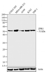
- Western blot analysis was performed on whole cell extracts (30 µg lysate) of COLO 205 (Lane 1), MDA-MB-231 (Lane 2), A549 (Lane 3), K562 (Lane4), THP-1 (Lane 5). The blots were probed with Anti- ZPR1 Mouse Monoclonal Antibody (Product # MA1-13003, 1:500 - 1:2000 dilution) and detected by chemiluminescence using Goat anti-Mouse IgG (H+L) Secondary Antibody, HRP conjugate (Product # 62-6520, 1:4000 dilution). A 51 kDa band corresponding to ZPR1 was observed across cell lines tested. Known quantity of protein samples were electrophoresed using Novex® NuPAGE® 4-12 % Bis-Tris gel (Product # NP0321BOX), XCell SureLock™ Electrophoresis System (Product # EI0002) and Novex® Sharp Pre-Stained Protein Standard (Product # LC5800). Resolved proteins were then transferred onto a transferred onto a nitrocellulose membrane with Pierce™ Power Blotter System (22834). The membrane was probed with the relevant primary and secondary Antibody following blocking with 5 % skimmed milk. Chemiluminescent detection was performed using Pierce™ ECL Western Blotting Substrate (Product # 32106).

Supportive validation
- Submitted by
- Invitrogen Antibodies (provider)
- Main image

- Experimental details
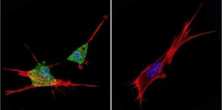
- Immunofluorescent analysis of ZPR1 in MCF-7 Cells. Cells were grown on chamber slides and fixed with formaldehyde prior to staining. Cells were probed without (control) or with a ZPR1 monoclonal antibody (Product # MA1-13003) at a dilution of 1:20 overnight at 4 C, washed with PBS and incubated with a DyLight-488 conjugated secondary antibody (Product # 35503). ZPR1 staining (green), F-Actin staining with Phalloidin (red) and nuclei with DAPI (blue) is shown. Images were taken at 60X magnification.

- Submitted by
- Invitrogen Antibodies (provider)
- Main image
- Experimental details
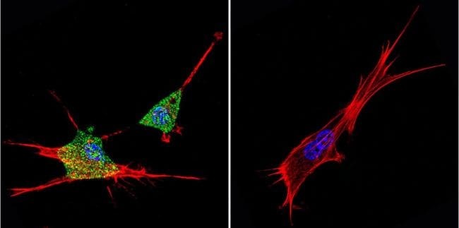
- Immunofluorescent analysis of ZPR1 in NIH-3T3 Cells. Cells were grown on chamber slides and fixed with formaldehyde prior to staining. Cells were probed without (control) or with a ZPR1 monoclonal antibody (Product # MA1-13003) at a dilution of 1:20 overnight at 4 C, washed with PBS and incubated with a DyLight-488 conjugated secondary antibody (Product # 35503). ZPR1 staining (green), F-Actin staining with Phalloidin (red) and nuclei with DAPI (blue) is shown. Images were taken at 60X magnification.
Supportive validation
- Submitted by
- Invitrogen Antibodies (provider)
- Main image
- Experimental details
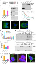
- Figure 2 Characterization of hSMN S290 phospho-mutant and phospho-mimetic. ( A ) Multiple sequence alignment of the C-termini of SMN from different species showing conservation of serine 290 residue. ( B , C ) Expression of hSMN-FL, hSMN S290D and hSMN S290A in NSC34 cells by Western blot 48 h or 72 h post-transfection (mean +- SEM, n = 6, one-way ANOVA with Sidak post-test, **** p < 0.0001). ( D , E ) Radioactive pulse-chase experiment determined stability of hSMN-FL, hSMN S290D and hSMN S290A. Representative autoradiographs at the same exposure times and decay curves of immunoprecipitated hSMN-FL, hSMN S290D, and hSMN S290A (mean +- SEM, n = 3, non-linear regression, one phase decay, R 2 (hSMN-FL) = 0.815, R 2 (hSMN S290D) = 0.854, R 2 (hSMN S290A) = 0.846). ( F ) Representative fluorescence images display NSC34 cells expressing GFP-tagged hSMN-FL, hSMN S290D or S290A. Scale bar represents 20 um. ( G ) Nuclear bodies (NBs) were quantified in NSC34 cells expressing hSMN-FL, hSMN S290D or hSMN S290A (mean +- SEM, n = 3, one-way ANOVA with Sidak post-test, ** p < 0.01). ( H , I ) NSC34 cells and ( J , K ) HEK 293T cells expressing GFP, GFP-tagged hSMN-FL, hSMN S290D and hSMN S290A ( H , I ) 48 h or ( J , K ) 24 h post-transfection. Co-immunoprecipitation of either ( H , I ) endogenous SMN and hSMN constructs or ( J , K ) ZPR1 and hSMN constructs were performed and determined by Western blot analysis. #, unspecific band. ( I ) Ratios of endogenous SMN to hSMN construct protein
