Explore
 Validate
 Learn
Learn Western blot
 ELISA
ELISAAntibody data
- Antibody Data
- Antigen structure
- References [0]
- Comments [0]
- Validations
- Western blot [1]
- Immunohistochemistry [1]
Submit
Validation data
Reference
Comment
Report error
- Product number
- AP09132PU-N - Provider product page

- Provider
- Acris Antibodies GmbH
- Proper citation
- Acris Antibodies GmbH Cat#AP09132PU-N, RRID:AB_2035390
- Product name
- anti Cyclin L1 (+ Cyclin L2, Isoform 2)
- Antibody type
- Polyclonal
- Antigen
- Prepared from whole rabbit serum produced by repeated immunizations with a synthetic construct consisting of full length Human Cyclin L1beta protein
- Reactivity
- Human
- Host
- Rabbit
- Isotype
- IgG
- Vial size
- 0.5 mg
- Concentration
- 5.0 mg/ml (by UV absorbance at 280 nm)
No comments: Submit comment
Supportive validation
- Submitted by
- Acris Antibodies GmbH (provider)
- Main image
- Experimental details
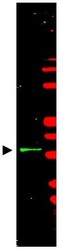
- Western blot using Protein A Purified anti-Cyclin L1/L2beta antibody shows detection of a band ~35 kDa corresponding to Cyclin L beta (arrowhead) present in mouse brain whole cell lysate (800 nm channel - green). Marker proteins appear red (700 nm channel) and were used for molecular weight comparisons. Approximately 35 µg of lysate was separated by 4-20% SDS-PAGE followed by transfer to nitrocellulose. After blocking the membrane was probed with the primary antibody diluted to 1:2,500 for 2h at room temperature followed by washes and reaction with a 1:10,000 dilution of IRDye(TM)800 conjugated Gt-a-Rabbit IgG [H&L] MX for 45 min at room temperature. IRDye(TM)800 fluorescence image was captured using the Odyssey(R) Infrared Imaging System developed by LI-COR. IRDye is a trademark of LI-COR, Inc. Other detection systems will yield similar results.

Supportive validation
- Submitted by
- Acris Antibodies GmbH (provider)
- Main image

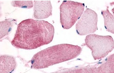
- Experimental details
- Immunohistochemistry. Affinity Purified anti- Cyclin L1/L2 (isoform 2) antibody was used at a 10 µg/ml to detect cyclin L in a variety of tissues including breast, kidney, liver, lung, skeletal muscle, pancreas, prostate and spleen. In some tissues elevated background staining was noted. In these instances further optimization of dilution is suggested. This image shows Cyclin L staining of human skeletal muscle. Tissue was formalinfixed and paraffin embedded.