Explore
 Validate
 Learn
Learn Western blot
 ELISA
ELISAAntibody data
- Antibody Data
- Antigen structure
- References [3]
- Comments [0]
- Validations
- Western blot [1]
- Immunocytochemistry [1]
- Immunohistochemistry [1]
- Flow cytometry [1]
Submit
Validation data
Reference
Comment
Report error
- Product number
- ABIN952698 - Provider product page

- Provider
- antibodies-online
- Product name
- anti-Histone Deacetylase 2 (HDAC2) (AA 417-447), (Middle Region) antibody
- Antibody type
- Polyclonal
- Antigen
- KLH conjugated synthetic peptide between 417~447 amino acids from the Central region of Human HDAC2
- Description
- Protein A column, followed by peptide affinity purification
- Reactivity
- Human
- Host
- Rabbit
- Epitope
- AA 417-447,Middle Region
- Vial size
- 0.4 mL
- Concentration
- 0.25 mg/mL
- Storage
- Store undiluted at 2-8°C for one month or (in aliquots) at -20°C for longer.
- Handling
- Avoid repeated freezing and thawing.
Submitted references HDAC inhibitors regulate claudin-1 expression in colon cancer cells through modulation of mRNA stability.
Protein acetylation in the cardiorenal axis: the promise of histone deacetylase inhibitors.
Transcriptional induction of MMP-10 by TGF-beta, mediated by activation of MEF2A and downregulation of class IIa HDACs.
Krishnan M, Singh AB, Smith JJ, Sharma A, Chen X, Eschrich S, Yeatman TJ, Beauchamp RD, Dhawan P
Oncogene 2010 Jan 14;29(2):305-12
Oncogene 2010 Jan 14;29(2):305-12
Protein acetylation in the cardiorenal axis: the promise of histone deacetylase inhibitors.
Bush EW, McKinsey TA
Circulation research 2010 Feb 5;106(2):272-84
Circulation research 2010 Feb 5;106(2):272-84
Transcriptional induction of MMP-10 by TGF-beta, mediated by activation of MEF2A and downregulation of class IIa HDACs.
Ishikawa F, Miyoshi H, Nose K, Shibanuma M
Oncogene 2010 Feb 11;29(6):909-19
Oncogene 2010 Feb 11;29(6):909-19
No comments: Submit comment
Supportive validation
- Submitted by
- antibodies-online (provider)
- Main image
- Experimental details
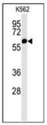
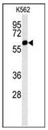
- Western blot analysis of HDAC2 Antibody (Center) Cat.-No AP52024PU-N in K562 cell line lysates (35ug/lane). HDAC2 (arrow) was detected using the purified Pab.
Supportive validation
- Submitted by
- antibodies-online (provider)
- Main image

- Experimental details
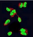
- Confocal immunofluorescent analysis of HDAC2 Antibody (Center) Cat.-No AP52024PU-N with 293 cell followed by Alexa Fluor 488-conjugated Goat anti-Rabbit lgG (green). Actin filaments have been labeled with Alexa Fluor 555 phalloidin (red).
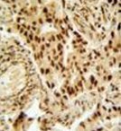
Supportive validation
- Submitted by
- antibodies-online (provider)
- Main image
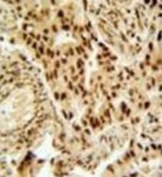
- Experimental details
- Immunohistochemistry analysis in formalin fixed and paraffin embedded human lung carcinoma reacted with HDAC2 Antibody (Center) Cat.-No AP52024PU-N, which was peroxidase conjugated to the secondary antibody and followed by DAB staining.
Supportive validation
- Submitted by
- antibodies-online (provider)
- Main image

- Experimental details
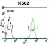
- Flow cytometric analysis of K562 cells using HDAC2 Antibody (Center) Cat.-No AP52024PU-N (right histogram) compared to a negative control cell (left histogram). FITC-conjugated goat-anti-rabbit secondary antibodies were used for the analysis.