Explore
 Validate
 Learn
Learn Western blot
 ELISA
ELISAAntibody data
- Antibody Data
- Antigen structure
- References [1]
- Comments [0]
- Validations
- Western blot [1]
- Immunoprecipitation [1]
Submit
Validation data
Reference
Comment
Report error
- Product number
- NB300-730 - Provider product page

- Provider
- Novus Biologicals
- Proper citation
- Novus Cat#NB300-730, RRID:AB_10001920
- Product name
- Rabbit Polyclonal HSF1 Antibody
- Antibody type
- Polyclonal
- Description
- Unpurified.
- Reactivity
- Human, Mouse, Rat
- Host
- Rabbit
- Isotype
- IgG
- Vial size
- 100uL
- Storage
- Store at -20C. Avoid freeze-thaw cycles.
Submitted references Deficiency in the Heat Stress Response Could Underlie Susceptibility to Metabolic Disease.
Rogers RS, Morris EM, Wheatley JL, Archer AE, McCoin CS, White KS, Wilson DR, Meers GM, Koch LG, Britton SL, Thyfault JP, Geiger PC
Diabetes 2016 Nov;65(11):3341-3351
Diabetes 2016 Nov;65(11):3341-3351
No comments: Submit comment
Supportive validation
- Submitted by
- Novus Biologicals (provider)
- Main image

- Experimental details
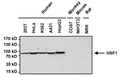
- Western Blot: HSF1 Antibody [NB300-730] - Analysis of Heat Shock Factor 1 (HSF1) was performed by loading 50ug of the indicated whole cell lysates per well onto a 4-20% Tris-HCl polyacrylamide gel. Proteins were transferred to a PVDF membrane and blocked with 5% BSA/TBST for at least 1 hour. The membrane was probed with a HSF1 rabbit polyclonal antibody at a dilution of 1:1000 overnight at 4C on a rocking platform, washed in TBS-0.1%Tween 20, and probed with a goat anti-rabbit IgG-HRP secondary antibody at a dilution of 1:20,000 for at least one hour. Chemiluminescent detection was performed using SuperSignal West Pico.
Supportive validation
- Submitted by
- Novus Biologicals (provider)
- Main image
- Experimental details
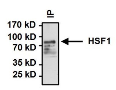
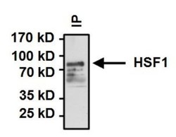
- Immunoprecipitation: HSF1 Antibody [NB300-730] - Analysis of Heat Shock Factor 1 (HSF 1) was performed on HeLa cells. Antigen: antibody complexes were formed by incubating 500ug whole cell lysate with 2ug of HSF1 polyclonal antibody overnight on a rocking platform at 4C. Immune complexes were captured on 50ul Protein A/G Plus Agarose, washed extensively, and eluted with 5X Lane Marker Reducing Sample Buffer. Samples were resolved on a 4-20% Tris-HCl polyacrylamide gel, transferred to a PVDF membrane, and blocked with 5% BSA/TBST for at least 1 hour. The membrane was probed with a HSF1 polyclonal antibody at a dilution of 1:1000 overnight rotating at 4C, washed in TBST, and probed with Clean Blot IP Detection Reagent-HRP at a dilution of 1:1000 for at least one hour. Chemiluminescent detection was performed using SuperSignal West Dura.