Explore
 Validate
 Learn
Learn Western blot
 ELISA
ELISAAntibody data
- Antibody Data
- Antigen structure
- References [2]
- Comments [0]
- Validations
- Western blot [1]
Submit
Validation data
Reference
Comment
Report error
- Product number
- A01044-2 - Provider product page

- Provider
- Boster Biological Technology
- Product name
- Anti-Caspase-7/CASP7 Antibody Picoband™
- Antibody type
- Polyclonal
- Description
- Rabbit IgG polyclonal antibody for Caspase-7/CASP7 detection. Tested with WB, IHC-P, ICC/IF, FCM, Direct ELISA in Human;Rat.
- Reactivity
- Human, Rat
- Host
- Rabbit
- Vial size
- 100μg/vial
- Concentration
- Add 0.2ml of distilled water will yield a concentration of 500ug/ml.
- Storage
- At -20°C for one year. After reconstitution, at 4°C for one month. It can also be aliquotted and stored frozen at -20°C for a longer time. Avoid repeated freezing and thawing.
- Handling
- Add 0.2ml of distilled water will yield a concentration of 500ug/ml.
Submitted references Isoflavones extracted from chickpea Cicer arietinum L. sprouts induce mitochondria-dependent apoptosis in human breast cancer cells.
Down-modulation of heat shock protein 70 and up-modulation of Caspase-3 during schisandrin B-induced apoptosis in human hepatoma SMMC-7721 cells.
Chen H, Ma HR, Gao YH, Zhang X, Habasi M, Hu R, Aisa HA
Phytotherapy research : PTR 2015 Feb;29(2):210-9
Phytotherapy research : PTR 2015 Feb;29(2):210-9
Down-modulation of heat shock protein 70 and up-modulation of Caspase-3 during schisandrin B-induced apoptosis in human hepatoma SMMC-7721 cells.
Wu YF, Cao MF, Gao YP, Chen F, Wang T, Zumbika EP, Qian KX
World journal of gastroenterology 2004 Oct 15;10(20):2944-8
World journal of gastroenterology 2004 Oct 15;10(20):2944-8
No comments: Submit comment
Supportive validation
- Submitted by
- Boster Biological Technology (provider)
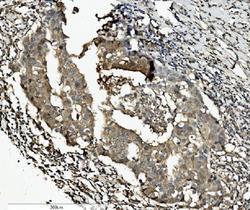
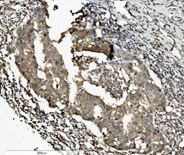
- Main image
- Experimental details
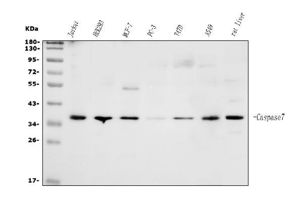
- Western blot analysis of Caspase-7/CASP7 using anti-Caspase-7/CASP7 antibody (A01044-2). Electrophoresis was performed on a 5-20% SDS-PAGE gel at 70V (Stacking gel) / 90V (Resolving gel) for 2-3 hours. The sample well of each lane was loaded with 30ug of sample under reducing conditions. Lane 1: human Jurkat whole cell lysates, Lane 2: human HEK293 whole cell lysates, Lane 3: human MCF-7 whole cell lysates, Lane 4: human PC-3 whole cell lysates, Lane 5: human T47D whole cell lysates, Lane 6: human A549 whole cell lysates, Lane 7: rat liver tissue lysates. After Electrophoresis, proteins were transferred to a Nitrocellulose membrane at 150mA for 50-90 minutes. Blocked the membrane with 5% Non-fat Milk/ TBS for 1.5 hour at RT. The membrane was incubated with rabbit anti-Caspase-7/CASP7 antigen affinity purified polyclonal antibody (Catalog # A01044-2) at 0.5 μg/mL overnight at 4°C, then washed with TBS-0.1%Tween 3 times with 5 minutes each and probed with a goat anti-rabbit IgG-HRP secondary antibody at a dilution of 1:5000 for 1.5 hour at RT. The signal is developed using an Enhanced Chemiluminescent detection (ECL) kit (Catalog # EK1002) with Tanon 5200 system. A specific band was detected for Caspase-7/CASP7 at approximately 35KD. The expected band size for Caspase-7/CASP7 is at 35KD.
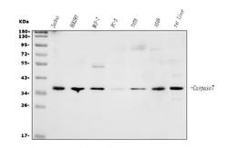
- Additional image