 Explore
 Validate
 Learn
Learn Western blot
Antibody data
- Antibody Data
- Antigen structure
- References [1]
- Comments [0]
- Validations
- Western blot [2]
- Immunohistochemistry [1]
Submit
Validation data
Reference
Comment
Report error
- Product number
- NB100-56529 - Provider product page

- Provider
- Novus Biologicals
- Proper citation
- Novus Cat#NB100-56529, RRID:AB_837863
- Product name
- Mouse Monoclonal Caspase-7 Antibody
- Antibody type
- Monoclonal
- Description
- Protein G purified. In Jurkat, a 35 kDa band is observed.
- Reactivity
- Human, Mouse, Rat
- Host
- Mouse
- Isotype
- IgG
- Vial size
- 0.1 mg
- Concentration
- 1.0 mg/ml
- Storage
- Store at 4C short term. Aliquot and store at -20C long term. Avoid freeze-thaw cycles.
Submitted references IER3 is a crucial mediator of TAp73β-induced apoptosis in cervical cancer and confers etoposide sensitivity.
Jin H, Suh DS, Kim TH, Yeom JH, Lee K, Bae J
Scientific reports 2015 Feb 10;5:8367
Scientific reports 2015 Feb 10;5:8367
No comments: Submit comment
Supportive validation
- Submitted by
- Novus Biologicals (provider)
- Main image
- Experimental details
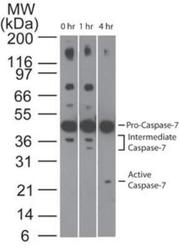
- Western Blot: Caspase-7 Antibody (25B881.1) [NB100-56529] - Analysis of Caspase-7 in Jurkat cells using Caspase-7 antibody at 1 ug/ml. Cells were treated with 2 uM staurosporine for different time periods. Caspase-7 activation is detected in western blots by the presence of Caspase-7 cleavage fragments. The antibody detected both pro (full-length) and active (cleaved) Caspase-7, depending on the treatment time points. A basal level of endogenous intermediate Caspase-7 can be see in untreated Jurkat cells.
- Submitted by
- Novus Biologicals (provider)
- Main image
- Experimental details
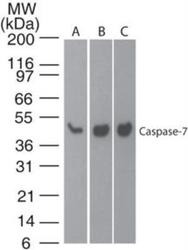
- Western Blot: Caspase-7 Antibody (25B881.1) [NB100-56529] - Analysis of Caspase-7 in A) human brain, B) mouse brain and C) rat brain lysate using Caspase-7 antibody at 1 ug/ml.

Supportive validation
- Submitted by
- Novus Biologicals (provider)
- Main image
- Experimental details
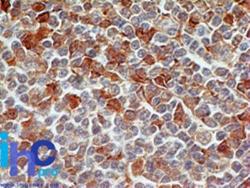
- Immunohistochemistry-Paraffin: Caspase-7 Antibody (25B881.1) [NB100-56529] - Human tonsil stained with Caspase-7 antibody at 5 ug/ml. Novus' human tonsil tissue slide was used for this test. Staining of formalin-fixed tissues is enhanced by boiling tissue sections in 10 mM sodium citrate buffer, pH 6.0 for 10-20 min followed by cooling at RT for 20 min.
