 Explore
 Validate
 Learn
LearnPA5-88517
antibody from Invitrogen Antibodies
Targeting: CGA
FSHA, GPA1, GPHa, GPHA1, HCG, LHA, TSHA
 Western blot
Antibody data
- Antibody Data
- Antigen structure
- References [1]
- Comments [0]
- Validations
- Western blot [2]
- Other assay [1]
Submit
Validation data
Reference
Comment
Report error
- Product number
- PA5-88517 - Provider product page

- Provider
- Invitrogen Antibodies
- Product name
- CGA Polyclonal Antibody
- Antibody type
- Polyclonal
- Antigen
- Recombinant full-length protein
- Description
- Immunogen sequence: APDVQDCPEC TLQENPFFSQ PGAPILQCMG CCFSRAYPTP LRSKKTMLVQ KNVTSESTCC VAKSYNRVTV MGGFKVENHT ACHCSTCYYH KS; Positive Samples: MCF7
- Reactivity
- Human
- Host
- Rabbit
- Isotype
- IgG
- Vial size
- 100 µL
- Concentration
- 0.23 mg/mL
- Storage
- -20° C, Avoid Freeze/Thaw Cycles
Submitted references Thyroid hormone receptor β sumoylation is required for thyrotropin regulation and thyroid hormone production.
Ke S, Liu YY, Karthikraj R, Kannan K, Jiang J, Abe K, Milanesi A, Brent GA
JCI insight 2021 Aug 23;6(16)
JCI insight 2021 Aug 23;6(16)
No comments: Submit comment
Supportive validation
- Submitted by
- Invitrogen Antibodies (provider)
- Main image

- Experimental details
- Western blot analysis of extracts of MCF-7 cells, using CGA Polyclonal antibody (Product # PA5-88517) at 1:1000 dilution. Secondary antibody: HRP Goat Anti-Rabbit IgG (H+L) at 1:10000 dilution. Lysates/proteins: 25ug per lane. Blocking buffer: 3% nonfat dry milk in TBST.

- Submitted by
- Invitrogen Antibodies (provider)
- Main image

- Experimental details
- Western Blot analysis of CGA in extracts of MCF-7 cells using CGA Polyclonal Antibody (Product # PA5-88517) at a dilution of 1:1000. A HRP Goat Anti-Rabbit IgG (H+L) secondary antibody was used at a dilution of 1:10,000. Lysates/proteins: 25 µg per lane. Blocking buffer: 3% nonfat dry milk in TBST.

Supportive validation
- Submitted by
- Invitrogen Antibodies (provider)
- Main image
- Experimental details
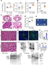
- Figure 3 Thyroid status, thyroid gland, and pituitary findings in THRB K146Q mice. ( A ) Serum T4, T3, and TSH concentrations in WT and THRB K146Q mice ( n = 13/ genotype) are shown as mean (+-SD) and paired t test for statistical analysis. TSH is shown in log 10 scale because of the wide differences in the levels in WT and K146Q mice. ( B ) Thyroid was rinsed with saline, patted dry, and weighed. The weight is shown as wet weight per mouse ( n = 18/genotype). ( C ) Representative histology of thyroid gland stained with H&E. Transverse section of thyroid gland (top panel) and thyroid follicles (lower panel). ( D ) Dissected pituitaries were rinsed with saline, patted dry, and weighed, and values are shown as wet weight of pituitary from each mouse ( n = 13/genotype). ( E ) Pituitaries are shown from WT and THRB K146Q mice. ( F ) Image of representative pituitary tissue histology with H&E stain from WT and THRB K146Q mutant mice. ( G ) Immunofluorescence staining for TSHbeta (green) and for nuclei (DAPI blue). Frozen sections of the pituitaries were incubated with anti-TSHbeta antibody at 1:50 dilution and conjugated with Alexa Fluor 488. ( H ) The TSHbeta-expressing cells and total cell numbers were counted using green and blue filters. ( I ) Western blot detection of TSHbeta and common glycoprotein alpha subunit (CGalpha) proteins. Pituitaries ( n = 3) were lysed in RIPA buffer, and 30 mug of protein was loaded on an 8% SDS gel. Membranes were Ponceau S-stained ( Supplementa
