Explore
 Validate
 Learn
Learn Western blot
Antibody data
- Antibody Data
- Antigen structure
- References [0]
- Comments [0]
- Validations
- Western blot [3]
- Immunocytochemistry [1]
- Immunohistochemistry [1]
Submit
Validation data
Reference
Comment
Report error
- Product number
- PA1-16507 - Provider product page

- Provider
- Invitrogen Antibodies
- Product name
- MTH1 Polyclonal Antibody
- Antibody type
- Polyclonal
- Antigen
- Synthetic peptide
- Description
- The target sequence has 91% sequence homology with mouse and rat. Suggested positive control: Hela whole cell extract, antigen standard for NUDT1 (transient overexpression lysate).
- Reactivity
- Human, Mouse, Rat
- Host
- Rabbit
- Isotype
- IgG
- Vial size
- 200 µL
- Concentration
- 1 mg/mL
- Storage
- -20° C, Avoid Freeze/Thaw Cycles
No comments: Submit comment
Supportive validation
- Submitted by
- Invitrogen Antibodies (provider)
- Main image

- Experimental details
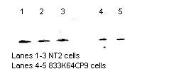
- Western Blot detection of MTH1 in NT2 cells and 833K54CP9 cells using Product # PA1-16507.
- Submitted by
- Invitrogen Antibodies (provider)
- Main image
- Experimental details
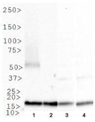
- Western blot analysis of MTH1 in 1. Ntera2, 2. MCF7 cell lysate, 3. A431 cell lysate and 4. COS7 cell lysate. Samples were incubated in MTH1 polyclonal antibody (Product # PA1-16507).

- Submitted by
- Invitrogen Antibodies (provider)
- Main image

- Experimental details
- Western blot analysis of MTH1 in 0.5 mg/mL A431 lysate. Samples were incubated in MTH1 polyclonal antibody (Product # PA1-16507). This experiment was performed under reducing conditions using the 12-230 kDa separation system.

Supportive validation
- Submitted by
- Invitrogen Antibodies (provider)
- Main image

- Experimental details
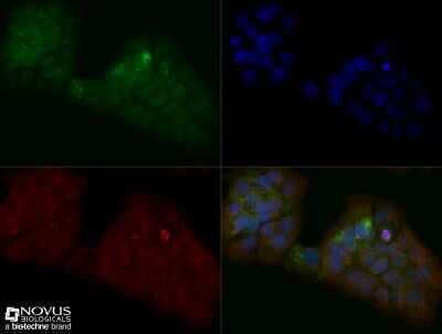
- Immunocytochemistry analysis of MTH1 in A431 cells fixed for 10 minutes using 10% formalin and then permeabilized for 5 minutes using 1X TBS + 0.5% Triton-X100. Samples were incubated in MTH1 polyclonal antibody (Product # PA1-16507) using a dilution of 1:200 dilution overnight at 4 °C followed by anti-rabbit DyLight 488 (Green) with a dilution of 1:500. Alpha tubulin was used as a co-stain at a 1:1000 dilution and detected with an anti-mouse DyLight 550 (Red) at a 1:500 dilution. Nuclei were counterstained with DAPI (Blue). Cells were imaged using a 40X objective.
Supportive validation
- Submitted by
- Invitrogen Antibodies (provider)
- Main image

- Experimental details
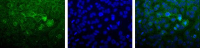
- Detection of MTH1 (Green) in Hela cells using Product # PA1-16507. Nuclei (Blue) are counterstained with Hoechst 33258.