Explore
 Validate
 Learn
Learn Western blot
 Immunoprecipitation
Antibody data
- Antibody Data
- Antigen structure
- References [0]
- Comments [0]
- Validations
- Western blot [2]
Submit
Validation data
Reference
Comment
Report error
- Product number
- PA1-29491 - Provider product page

- Provider
- Invitrogen Antibodies
- Product name
- LHX3 Polyclonal Antibody
- Antibody type
- Polyclonal
- Antigen
- Other
- Description
- Predicted molecular weight of 44 kDa.
- Reactivity
- Human, Mouse, Rat
- Host
- Rabbit
- Isotype
- IgG
- Vial size
- 50 µg
- Concentration
- 1 mg/mL
- Storage
- Store at 4°C short term. For long term storage, store at -20°C, avoiding freeze/thaw cycles.
No comments: Submit comment
Supportive validation
- Submitted by
- Invitrogen Antibodies (provider)
- Main image

- Experimental details
- Western blot was performed using Anti-LHX3 Rabbit Polyclonal Antibody (Product # PA1-29491) and a 44 kDa band corresponding to LHX3 was observed in Mouse Brain and Cerebellum tissue lysates but not in other tissues which are reported to be negative. Tissue extracts (30 µg lysate) of Mouse brain (Lane 1), Mouse cerebellum (Lane 2), Mouse liver (Lane 3) and Mouse kidney (Lane 4) were electrophoresed using Novex® NuPAGE® 12 % Bis-Tris gel (Product # NP0342BOX). Resolved proteins were then transferred onto a nitrocellulose membrane (Product #) by iBlot® 2 Dry Blotting System (Product # IB21001). The bot was probed with the primary antibody (1 µg/mL) and detected by chemiluminescence with Goat anti-Rabbit IgG (H+L) Superclonal™ Recombinant Secondary Antibody, HRP (Product # A27036, 1:4000 dilution) using the iBright FL 1000 (Product # A32752). Chemiluminescent detection was performed using Novex® ECL Chemiluminescent Substrate Reagent Kit (Product # WP20005).

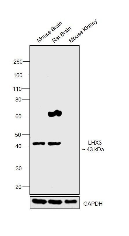
- Submitted by
- Invitrogen Antibodies (provider)
- Main image

- Experimental details
- Western blot was performed using Anti-LHX3 Polyclonal Antibody (Product # PA1-29491) and a 43 kDa band corresponding to LHX3 was observed across tissues tested except Mouse Kidney which is reported to be negative. Tissue extracts (30 µg lysate) of Mouse Brain (Lane 1), Rat Brain (Lane 2) and Mouse Kidney (Lane 3) were electrophoresed using NuPAGE™ 4-12% Bis-Tris Protein Gel (Product # NP0322BOX). Resolved proteins were then transferred onto a nitrocellulose membrane (Product # IB23001) by iBlot® 2 Dry Blotting System (Product # IB21001). The blot was probed with the primary antibody (1:2500 dilution) and detected by chemiluminescence with Goat anti-Rabbit IgG (H+L) Superclonal™ Recombinant Secondary Antibody, HRP (Product # A27036, 1:4000 dilution) using the iBright FL 1000 (Product # A32752). Chemiluminescent detection was performed using SuperSignal™ West Dura Extended Duration Substrate (Product # 34076).