 Explore
 Validate
 Learn
Learn Western blot
 Immunohistochemistry
Antibody data
- Antibody Data
- Antigen structure
- References [1]
- Comments [0]
- Validations
- Western blot [1]
- Immunocytochemistry [1]
- Immunohistochemistry [12]
Submit
Validation data
Reference
Comment
Report error
- Product number
- HPA008461 - Provider product page

- Provider
- Atlas Antibodies
- Proper citation
- Atlas Antibodies Cat#HPA008461, RRID:AB_1080461
- Product name
- Anti-SUN1
- Antibody type
- Polyclonal
- Reactivity
- Human
- Host
- Rabbit
- Conjugate
- Unconjugated
- Antigen sequence
QGNFFSFLPVLNWASMHRTQRVDDPQDVFKPTTSR
LKQPLQGDSEAFPWHWMSGVEQQVASLSGQCHHHG
ENLRELTTLLQKLQARVDQMEGGAAGPSASVRDAV
GQPPRETDFMAFHQEHEVRMSHLEDILGKLR- Isotype
- IgG
- Vial size
- 100 µl
- Storage
- Store at +4°C for short term storage. Long time storage is recommended at -20°C.
Submitted references Nuclear envelope-associated endosomes deliver surface proteins to the nucleus
Chaumet A, Wright G, Seet S, Tham K, Gounko N, Bard F
Nature Communications 2015 September;6
Nature Communications 2015 September;6
No comments: Submit comment
Supportive validation
- Submitted by
- Atlas Antibodies (provider)
- Main image
- Experimental details
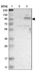
- Lane 1: Marker [kDa] 230, 130, 95, 72, 56, 36, 28, 17, 11Lane 2: Human cell line RT-4Lane 3: Human cell line U-251MG sp

Supportive validation
- Submitted by
- Atlas Antibodies (provider)
- Main image

- Experimental details
- Immunofluorescent staining of human cell line U-2 OS shows localization to nuclear membrane & vesicles.
- Sample type
- HUMAN

Enhanced validation
Enhanced validation
Supportive validation
- Submitted by
- Atlas Antibodies (provider)
- Enhanced method
- Orthogonal validation
- Main image

- Experimental details
- Immunohistochemistry analysis in human skin and bone marrow tissues using HPA008461 antibody. Corresponding SUN1 RNA-seq data are presented for the same tissues.
- Sample type
- HUMAN

Enhanced validation
- Submitted by
- Atlas Antibodies (provider)
- Enhanced method
- Independent antibody validation
- Main image

- Experimental details
- Immunohistochemical staining of human fallopian tube, kidney, skin and testis using Anti-SUN1 antibody HPA008461 (A) shows similar protein distribution across tissues to independent antibody HPA008346 (B).

Supportive validation
- Submitted by
- Atlas Antibodies (provider)
- Main image

- Experimental details
- Immunohistochemical staining of human placenta shows strong nuclear membrane positivity in trophoblastic cells.

- Submitted by
- Atlas Antibodies (provider)
- Main image
- Experimental details
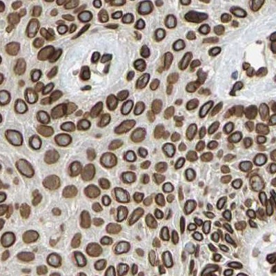
- Immunohistochemical staining of human skin using Anti-SUN1 antibody HPA008461.
- Sample type
- HUMAN
- Submitted by
- Atlas Antibodies (provider)
- Main image

- Experimental details
- Immunohistochemical staining of human pancreas using Anti-SUN1 antibody HPA008461.
- Sample type
- HUMAN

- Submitted by
- Atlas Antibodies (provider)
- Main image

- Experimental details
- Immunohistochemical staining of human lymph node using Anti-SUN1 antibody HPA008461.
- Sample type
- HUMAN
- Submitted by
- Atlas Antibodies (provider)
- Main image

- Experimental details
- Immunohistochemical staining of human colon using Anti-SUN1 antibody HPA008461.
- Sample type
- HUMAN

- Submitted by
- Atlas Antibodies (provider)
- Main image

- Experimental details
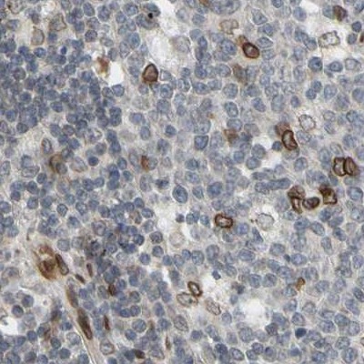
- Immunohistochemical staining of human bone marrow shows low positivity in hematopoietic cells as expected.
- Sample type
- HUMAN

- Submitted by
- Atlas Antibodies (provider)
- Main image
- Experimental details
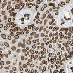
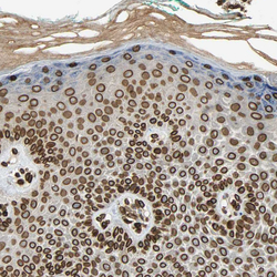
- Immunohistochemical staining of human skin shows strong positivity in nuclear membrane in squamous epithelial cells.
- Sample type
- HUMAN

- Submitted by
- Atlas Antibodies (provider)
- Main image

- Experimental details
- Immunohistochemical staining of human Fallopian tube shows moderate positivity in nuclear membrane in glandular cells.
- Sample type
- HUMAN

- Submitted by
- Atlas Antibodies (provider)
- Main image

- Experimental details
- Immunohistochemical staining of human testis shows strong positivity in nuclear membrane in Leydig cells.
- Sample type
- HUMAN

- Submitted by
- Atlas Antibodies (provider)
- Main image

- Experimental details
- Immunohistochemical staining of human kidney shows strong positivity in nuclear membrane in cells in glomeruli.
- Sample type
- HUMAN
