Explore
 Validate
 Learn
Learn Western blot
 Immunocytochemistry
 Immunohistochemistry
Antibody data
- Antibody Data
- Antigen structure
- References [1]
- Comments [0]
- Validations
- Immunocytochemistry [1]
- Immunohistochemistry [4]
Submit
Validation data
Reference
Comment
Report error
- Product number
- HPA012072 - Provider product page

- Provider
- Atlas Antibodies
- Proper citation
- Atlas Antibodies Cat#HPA012072, RRID:AB_1852714
- Product name
- Anti-LASP1
- Antibody type
- Polyclonal
- Reactivity
- Human, Mouse, Rat
- Host
- Rabbit
- Conjugate
- Unconjugated
- Antigen sequence
ADTPENLRLKQQSELQSQVRYKEEFEKNKGKGFSV
VADTPELQRIKKTQDQISNIKYHEEFEKSRMGPSG
GEGMEPERRDSQDGSSYRRPLEQQQPHHIPTSAPV
YQQPQQQPVAQSYGGYKE- Isotype
- IgG
- Vial size
- 100 µl
- Storage
- Store at +4°C for short term storage. Long time storage is recommended at -20°C.
Submitted references A complex containing LPP and α-actinin mediates TGFβ-induced migration and invasion of ErbB2-expressing breast cancer cells.
Ngan E, Northey JJ, Brown CM, Ursini-Siegel J, Siegel PM
Journal of cell science 2013 May 1;126(Pt 9):1981-91
Journal of cell science 2013 May 1;126(Pt 9):1981-91
No comments: Submit comment
Supportive validation
- Submitted by
- Atlas Antibodies (provider)
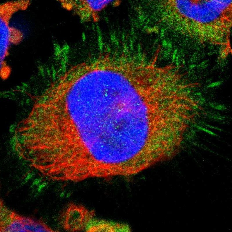
- Main image

- Experimental details
- Immunofluorescent staining of human cell line U-251 MG shows localization to plasma membrane, cytosol & focal adhesion sites.
- Sample type
- HUMAN
Enhanced validation
Supportive validation
- Submitted by
- Atlas Antibodies (provider)
- Enhanced method
- Orthogonal validation
- Main image
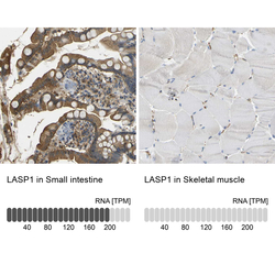
- Experimental details
- Immunohistochemistry analysis in human small intestine and skeletal muscle tissues using Anti-LASP1 antibody. Corresponding LASP1 RNA-seq data are presented for the same tissues.
- Sample type
- HUMAN

Supportive validation
- Submitted by
- Atlas Antibodies (provider)
- Main image
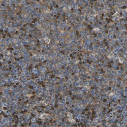
- Experimental details
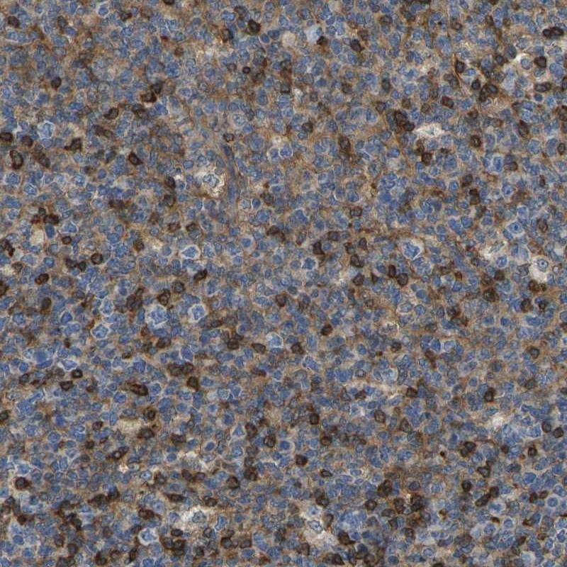
- Immunohistochemical staining of human tonsil shows distinct cytoplasmic positivity in cells in reaction center.
- Submitted by
- Atlas Antibodies (provider)
- Main image

- Experimental details
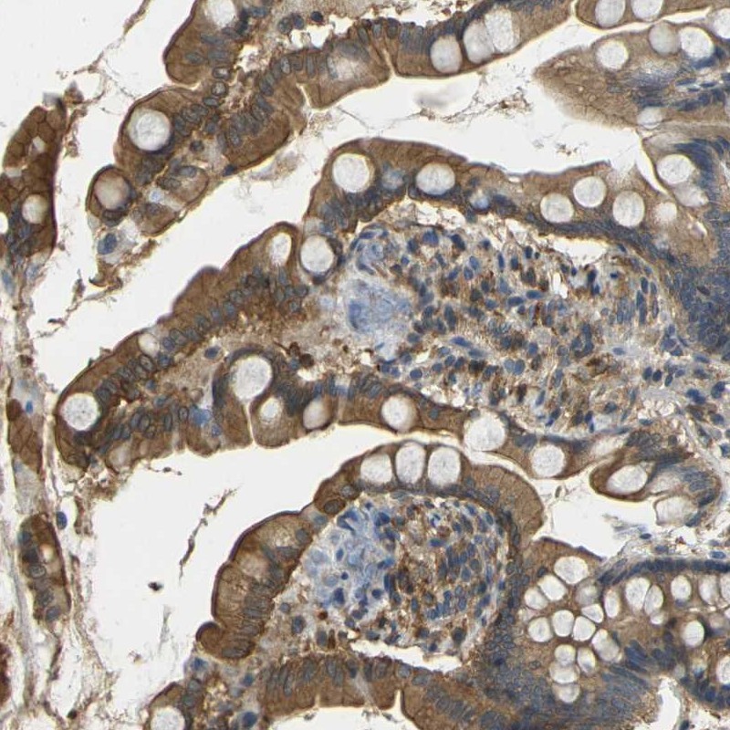
- Immunohistochemical staining of human small intestine shows high expression.
- Sample type
- HUMAN
- Submitted by
- Atlas Antibodies (provider)
- Main image
- Experimental details
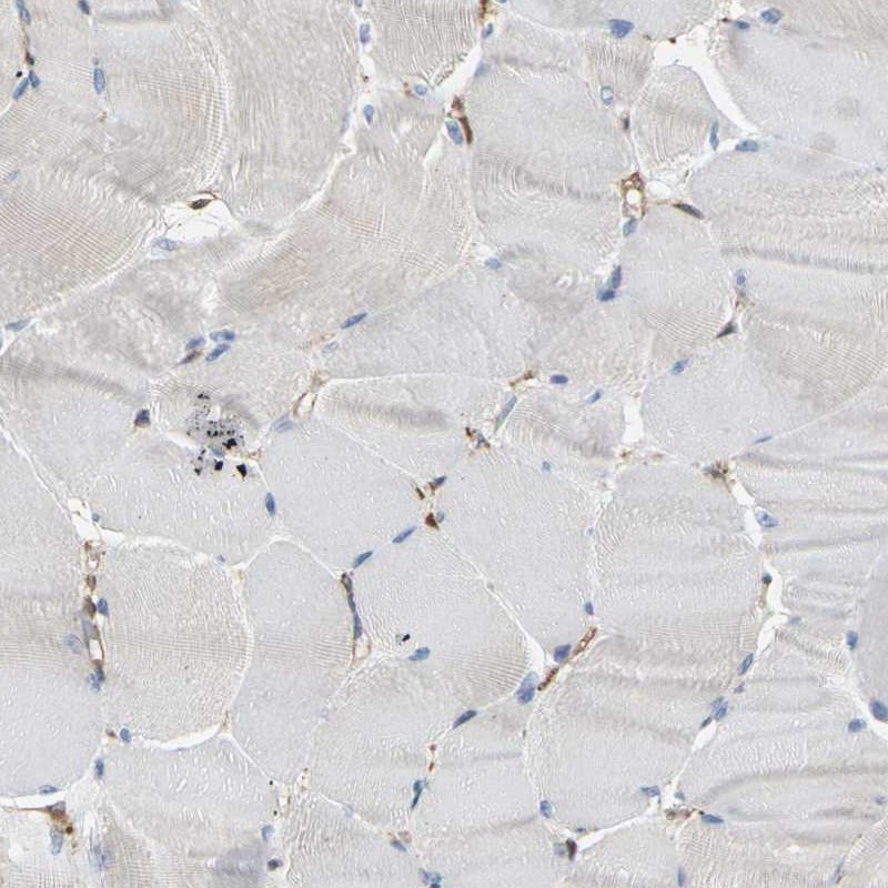
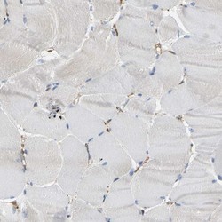
- Immunohistochemical staining of human skeletal muscle shows low expression as expected.
- Sample type
- HUMAN