Explore
 Validate
 Learn
Learn Western blot
 Immunoprecipitation
Antibody data
- Antibody Data
- Antigen structure
- References [0]
- Comments [0]
- Validations
- Western blot [6]
- Immunocytochemistry [2]
- Immunohistochemistry [1]
- Other assay [1]
Submit
Validation data
Reference
Comment
Report error
- Product number
- PA5-28937 - Provider product page

- Provider
- Invitrogen Antibodies
- Product name
- 14-3-3 epsilon Polyclonal Antibody
- Antibody type
- Polyclonal
- Antigen
- Recombinant full-length protein
- Description
- Recommended positive controls: 293T, A431, HeLa, HepG2, Mouse brain. Predicted reactivity: Mouse (100%), Rat (98%), Xenopus laevis (96%), Chicken (100%), Chimpanzee (100%), Bovine (100%). Store product as a concentrated solution. Centrifuge briefly prior to opening the vial.
- Reactivity
- Human, Mouse, Rat
- Host
- Rabbit
- Isotype
- IgG
- Vial size
- 100 µL
- Concentration
- 0.91 mg/mL
- Storage
- Store at 4°C short term. For long term storage, store at -20°C, avoiding freeze/thaw cycles.
No comments: Submit comment
Supportive validation
- Submitted by
- Invitrogen Antibodies (provider)
- Main image
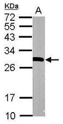
- Experimental details
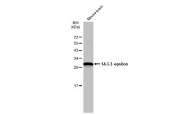
- Western blot analysis of 14-3-3 epsilon using 20 µg of mouse brain lysate. Samples were loaded onto a 12% SDS-PAGE gel and probed with a 14-3-3 epsilon polyclonal antibody (Product # PA5-28937) at a dilution of 1:10,000.

- Submitted by
- Invitrogen Antibodies (provider)
- Main image
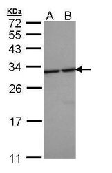
- Experimental details
- Western blot analysis of 14-3-3 epsilon using 30 µg of A) H1299 and B) HeLa S3 lysate. Samples were loaded onto a 12% SDS-PAGE gel and probed with a 14-3-3 epsilon polyclonal antibody (Product # PA5-28937) at a dilution of 1:5000.
- Submitted by
- Invitrogen Antibodies (provider)
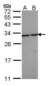
- Main image
- Experimental details
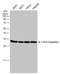
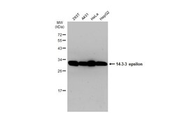
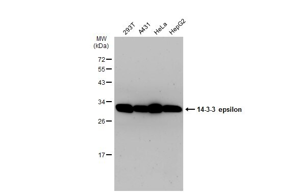
- Western Blot analysis of 14-3-3-epsilon was performed by separating 30 µg of various whole cell extracts by 12% SDS-PAGE. Proteins were transferred to a membrane and probed with a 14-3-3 epsilon Polyclonal Antibody (Product # PA5-28937) at a dilution of 1:5000 and a HRP-conjugated anti-rabbit IgG secondary antibody.
- Submitted by
- Invitrogen Antibodies (provider)
- Main image
- Experimental details
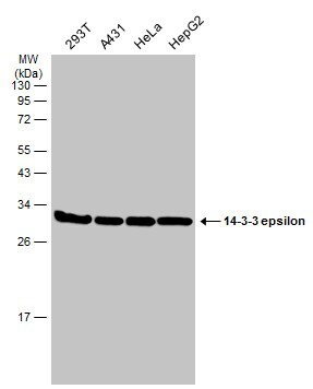
- Western Blot using 14-3-3 epsilon Polyclonal Antibody (Product # PA5-28937). Various whole cell extracts (30 µg) were separated by 12% SDS-PAGE, and the membrane was blotted with 14-3-3 epsilon Polyclonal Antibody (Product # PA5-28937) diluted at 1:5,000. The HRP-conjugated anti-rabbit IgG antibody was used to detect the primary antibody.
- Submitted by
- Invitrogen Antibodies (provider)
- Main image
- Experimental details
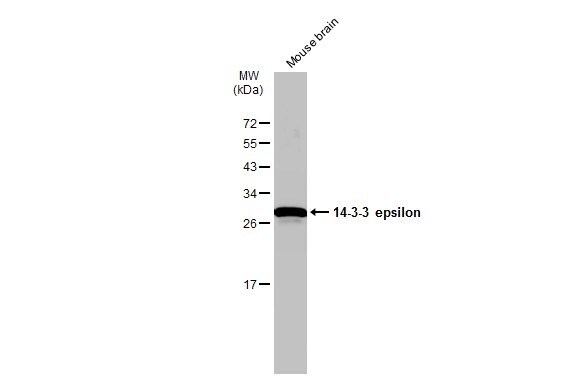
- Western Blot using 14-3-3 epsilon Polyclonal Antibody (Product # PA5-28937). Mouse tissue extract (50 µg) was separated by 12% SDS-PAGE, and the membrane was blotted with 14-3-3 epsilon Polyclonal Antibody (Product # PA5-28937) diluted at 1:6,000. The HRP-conjugated anti-rabbit IgG antibody was used to detect the primary antibody.
- Submitted by
- Invitrogen Antibodies (provider)
- Main image
- Experimental details
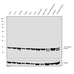
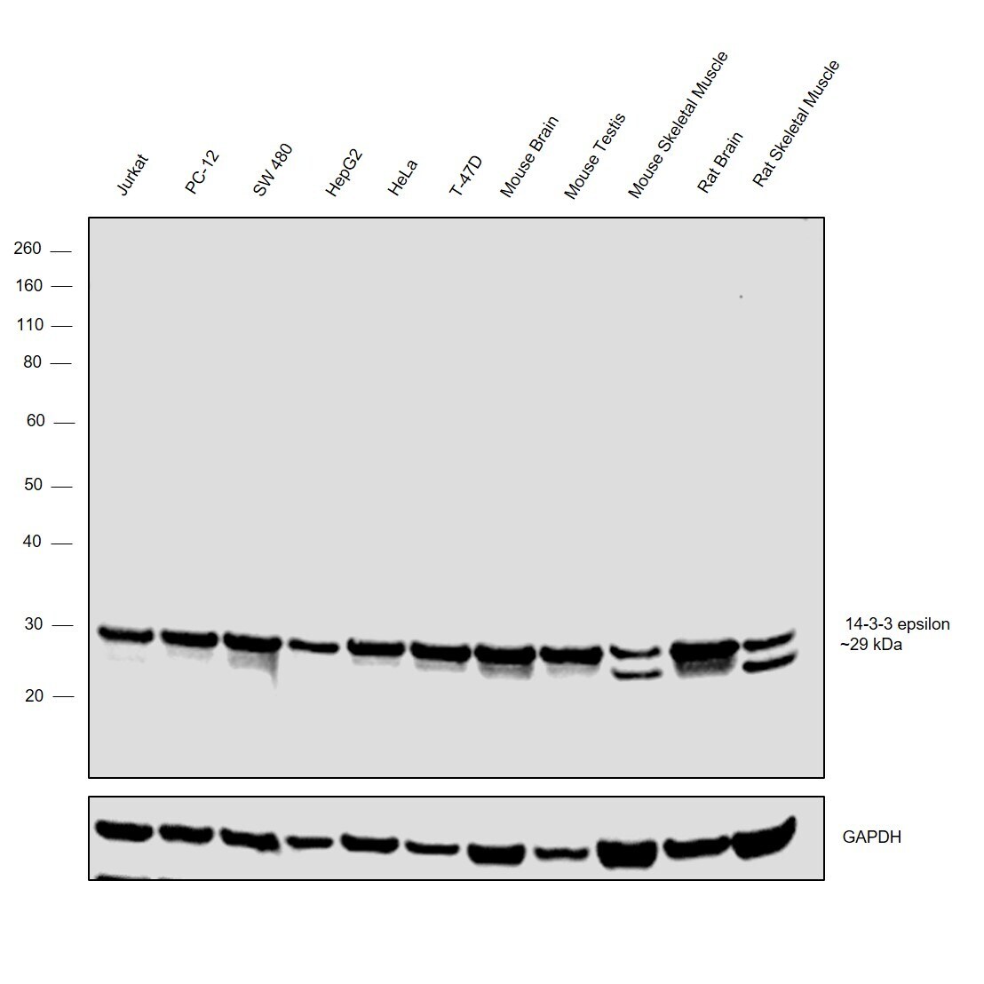
- Western blot was performed using Anti-14-3-3 epsilon Polyclonal Antibody (Product # PA5-28937) and a ~29 kDa band was observed across the panel tested. Whole cell extracts (30 µg lysate) of Jurkat (Lane 1), PC-12 (Lane 2), SW 480 (Lane 3), HepG2 (Lane 4), HeLa (Lane 5), T-47D (Lane 6) and tissue extracts (30 µg lysate) of Mouse Brain (Lane 7), Mouse Testis (Lane 8), Mouse Skeletal Muscle (Lane 9), Rat Brain (Lane 10) and Rat Skeletal Muscle (Lane 11) were electrophoresed using NuPAGE™ 4-12% Bis-Tris Protein Gel (Product # NP0321BOX). Resolved proteins were then transferred onto a nitrocellulose membrane (Product # IB23001) by iBlot® 2 Dry Blotting System (Product # IB21001). The blot was probed with the primary antibody (1:1000 dilution) and detected by chemiluminescence with Goat anti-Rabbit IgG (H+L), Superclonal™ Recombinant Secondary Antibody, HRP (Product # A27036, 1:4000 dilution) using the iBright FL 1000 (Product # A32752). Chemiluminescent detection was performed using Novex® ECL Chemiluminescent Substrate Reagent Kit (Product # WP20005).
Supportive validation
- Submitted by
- Invitrogen Antibodies (provider)
- Main image

- Experimental details
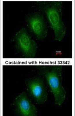
- Immunofluorescent analysis of 14-3-3 epsilon in methanol-fixed HeLa cells using a 14-3-3 epsilon polyclonal antibody (Product # PA5-28937) at a 1:200 dilution.
- Submitted by
- Invitrogen Antibodies (provider)
- Main image

- Experimental details
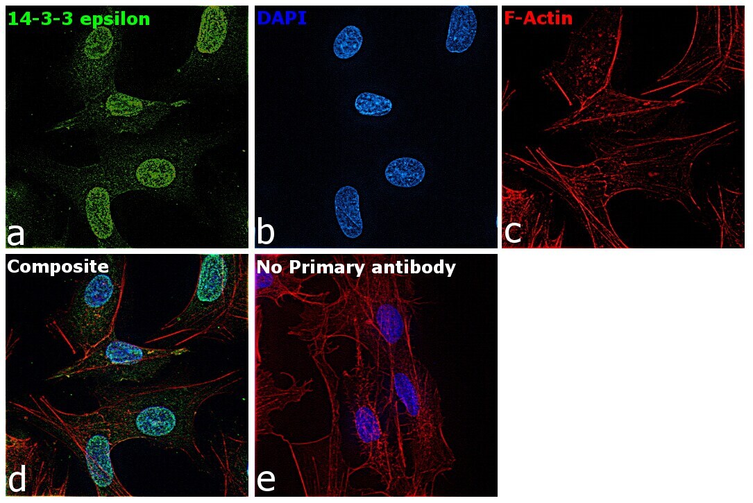
- Immunofluorescence analysis of 14-3-3 epsilon was performed using Hep G2 cells. The cells were fixed with 4% paraformaldehyde for 10 minutes, permeabilized with 0.1% Triton™ X-100 for 15 minutes, and blocked with 2% BSA for 1 hour at room temperature. The cells were labeled with 14-3-3 epsilon Polyclonal Antibody (Product # PA5-28937) at 1:200 dilution in 0.1% BSA and incubated overnight at 4 degree and then labeled with Goat anti-Rabbit IgG (H+L) Superclonal™ Recombinant Secondary Antibody, Alexa Fluor® 488 (Product # A27034, 1:2000 dilution) for 45 minutes at room temperature (Panel a: green) in HeLa cells. Nuclei (Panel b: blue) were stained with ProLong™ Diamond Antifade Mountant with DAPI (Product # P36962). F-actin (Panel c: red) was stained with Rhodamine Phalloidin (Product # R415, 1:300). Panel d represents the merged image of Hep G2 cells showing nuclear and cytoplasmic localization for 14-3-3 epsilon. Panel e represents control cells with no primary antibody to assess background. The images were captured at 60X magnification.
Supportive validation
- Submitted by
- Invitrogen Antibodies (provider)
- Main image
- Experimental details
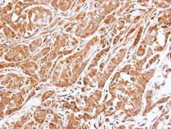
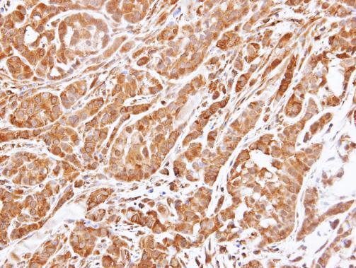
- Immunohistochemical analysis of paraffin-embedded A549 xenograft , using 14-3-3 epsilon (Product # PA5-28937) antibody at 1:500 dilution. Antigen Retrieval: EDTA based buffer, pH 8.0, 15 min.
Supportive validation
- Submitted by
- Invitrogen Antibodies (provider)
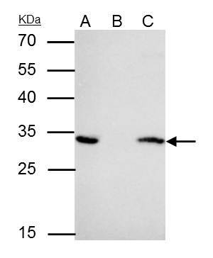
- Main image
- Experimental details
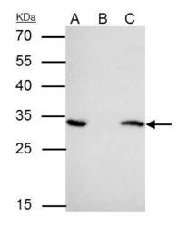
- 14-3-3 epsilon Polyclonal Antibody immunoprecipitates YWHAE protein in IP experiments. IP samples: HeLa whole cell extract. A. 40 µg HeLa whole cell extract. B. Control with 4 µg of preimmune Rabbit IgG. C. Immunoprecipitation of YWHAE protein by 4 µg 14-3-3 epsilon Polyclonal Antibody (Product # PA5-28937). 12 % SDS-PAGE. The immunoprecipitated YWHAE protein was detected by 14-3-3 epsilon Polyclonal Antibody (Product # PA5-28937) diluted at 1:1,000.