Explore
 Validate
 Learn
Learn Western blot
Antibody data
- Antibody Data
- Antigen structure
- References [0]
- Comments [0]
- Validations
- Western blot [1]
- Immunocytochemistry [4]
- Immunohistochemistry [2]
- Flow cytometry [2]
Submit
Validation data
Reference
Comment
Report error
- Product number
- PA5-140980 - Provider product page

- Provider
- Invitrogen Antibodies
- Product name
- FPR1 Polyclonal Antibody
- Antibody type
- Polyclonal
- Antigen
- Synthetic peptide
- Reactivity
- Human, Mouse
- Host
- Rabbit
- Isotype
- IgG
- Vial size
- 100 µL
- Concentration
- 1 mg/mL
- Storage
- Store at 4°C short term. For long term storage, store at -20°C, avoiding freeze/thaw cycles.
No comments: Submit comment
Supportive validation
- Submitted by
- Invitrogen Antibodies (provider)
- Main image

- Experimental details
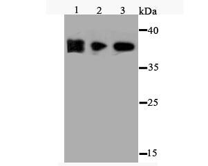
- Western blot analysis of FPR1 in different cell lysates. Samples were incubated in FPR1 Polyclonal antibody (Product # PA5-140980) using a dilution of 1:500. Positive control: Lane 1: NCCIT; Lane 2: MEF; Lane 3: HES.
Supportive validation
- Submitted by
- Invitrogen Antibodies (provider)
- Main image

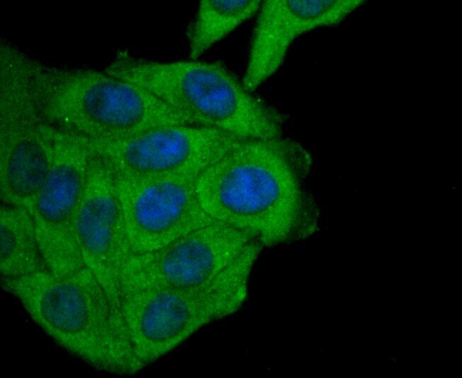
- Experimental details
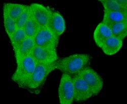
- Immunocytochemistry analysis of FPR1 in MCF-7 cells (green). Samples were incubated in FPR1 Polyclonal antibody (Product # PA5-140980). The nuclear counter stain is DAPI (blue). Cells were fixed in paraformaldehyde, permeabilized with 0.25% Triton X100/PBS.

- Submitted by
- Invitrogen Antibodies (provider)
- Main image
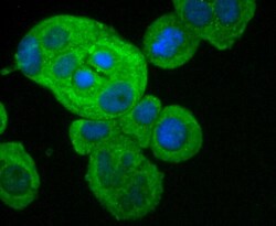
- Experimental details
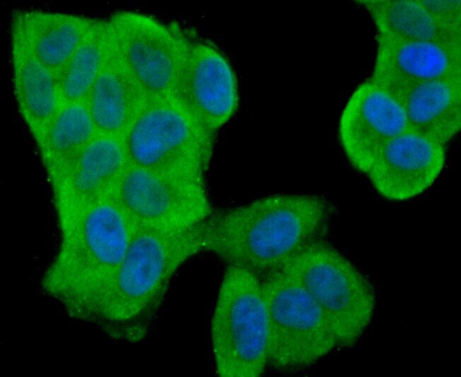
- Immunocytochemistry analysis of FPR1 in HeLa cells (green). Samples were incubated in FPR1 Polyclonal antibody (Product # PA5-140980). The nuclear counter stain is DAPI (blue). Cells were fixed in paraformaldehyde, permeabilized with 0.25% Triton X100/PBS.
- Submitted by
- Invitrogen Antibodies (provider)
- Main image

- Experimental details
- Immunocytochemistry analysis of FPR1 in HepG2 cells (green). Samples were incubated in FPR1 Polyclonal antibody (Product # PA5-140980). The nuclear counter stain is DAPI (blue). Cells were fixed in paraformaldehyde, permeabilized with 0.25% Triton X100/PBS.
- Submitted by
- Invitrogen Antibodies (provider)
- Main image
- Experimental details
- Immunocytochemistry analysis of FPR1 in MCF-7 cells (green). Samples were incubated in FPR1 Polyclonal antibody (Product # PA5-140980). The nuclear counter stain is DAPI (blue). Cells were fixed in paraformaldehyde, permeabilized with 0.25% Triton X100/PBS.

Supportive validation
- Submitted by
- Invitrogen Antibodies (provider)
- Main image
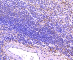
- Experimental details
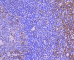
- Immunohistochemistry analysis of FPR1 in paraffin-embedded human spleen tissue. Samples were incubated in FPR1 Polyclonal antibody (Product # PA5-140980). Counter stained with hematoxylin.
- Submitted by
- Invitrogen Antibodies (provider)
- Main image
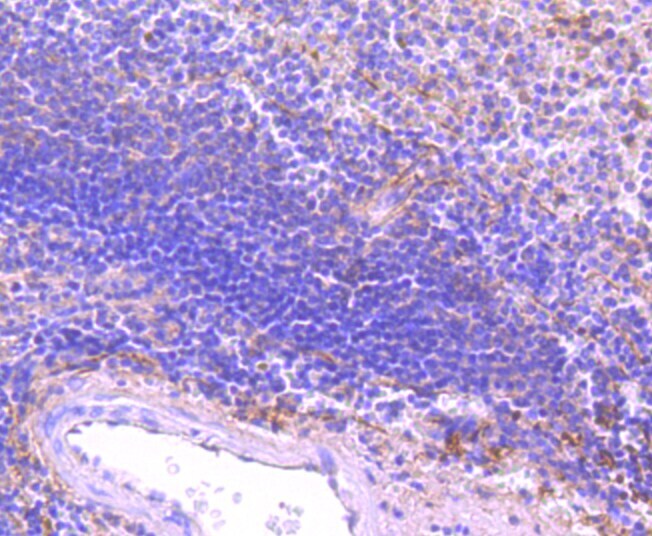
- Experimental details
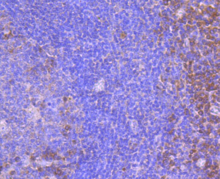
- Immunohistochemistry analysis of FPR1 in paraffin-embedded human tonsil tissue. Samples were incubated in FPR1 Polyclonal antibody (Product # PA5-140980). Counter stained with hematoxylin.
Supportive validation
- Submitted by
- Invitrogen Antibodies (provider)
- Main image
- Experimental details
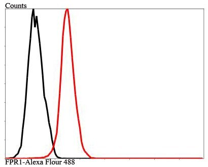
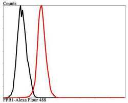
- Flow cytometry of FPR1 in MCF-7 cells. Samples were incubated in FPR1 Polyclonal antibody (Product # PA5-140980) using a dilution of 1:100 (red). Unlabeled control (cells without incubation with primary antibody; black).

- Submitted by
- Invitrogen Antibodies (provider)
- Main image
- Experimental details
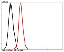
- Flow cytometry of FPR1 in MCF-7 cells. Samples were incubated in FPR1 Polyclonal antibody (Product # PA5-140980) using a dilution of 1:100 (red). Unlabeled control (cells without incubation with primary antibody; black).