Antibody data
- Antibody Data
- Antigen structure
- References [1]
- Comments [0]
- Validations
- Western blot [3]
- Immunocytochemistry [3]
Submit
Validation data
Reference
Comment
Report error
- Product number
- PA5-35383 - Provider product page

- Provider
- Invitrogen Antibodies
- Product name
- alpha-II Spectrin Polyclonal Antibody
- Antibody type
- Polyclonal
- Antigen
- Recombinant full-length protein
- Description
- This antibody contains enough material to conduct 10 mini-Western blots.
- Reactivity
- Human, Mouse, Rat
- Host
- Rabbit
- Isotype
- IgG
- Vial size
- 100 µL
- Storage
- -20° C, Avoid Freeze/Thaw Cycles
Submitted references Retinoic acid degradation shapes zonal development of vestibular organs and sensitivity to transient linear accelerations.
Ono K, Keller J, López Ramírez O, González Garrido A, Zobeiri OA, Chang HHV, Vijayakumar S, Ayiotis A, Duester G, Della Santina CC, Jones SM, Cullen KE, Eatock RA, Wu DK
Nature communications 2020 Jan 2;11(1):63
Nature communications 2020 Jan 2;11(1):63
No comments: Submit comment
Supportive validation
- Submitted by
- Invitrogen Antibodies (provider)
- Main image
- Experimental details
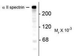
- Western blot analysis of Alpha II Spectrin in mouse brain lysate using a Alpha II Spectrin polyclonal antibody (Product # PA5-35383). Results show a band at ~240kDa.
- Submitted by
- Invitrogen Antibodies (provider)
- Main image
- Experimental details
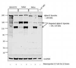
- Western blot analysis was performed on membrane enriched extracts (30 µg lysate) of SH-SY5Y (Lane 1), SH-SY5Y treated with Staurosporine (0.5 uM for 24 hours) (Lane 2), Jurkat (Lane 3), Jurkat treated with Staurosporine (0.5 uM for 24 hours) (Lane 4), HeLa (Lane 5), HeLa treated with Staurosporine (0.5 uM for 24 hours) (Lane 6) and U-87 MG (Lane 7). The blot was probed with Anti-alpha-II Spectrin Polyclonal Antibody (Product # PA5-35383, 1:5000 dilution) and detected by chemiluminescence using Goat anti-Rabbit IgG (H+L) Superclonal™ Secondary Antibody, HRP conjugate (Product # A27036, 0.25 µg/mL, 1:4000 dilution). A 260 kDa band corresponding to intact alpha-II Spectrin was observed across the untreated lysates tested. Upon Staurosporine treatment, alpha-II Spectrin degrades into two major fragments at 160 and 120 kDa.
- Submitted by
- Invitrogen Antibodies (provider)
- Main image
- Experimental details
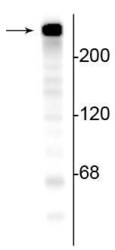
- Western blot of SPTAN1 in mouse brain lysate showing specific immunolabeling of a band at ~240 kDa corresponding to alpha-II Spectrin polyclonal antibody (Product # PA5-35383).
Supportive validation
- Submitted by
- Invitrogen Antibodies (provider)
- Main image
- Experimental details
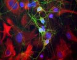
- Immunofluorescent analysis of Alpha II Spectrin in cultured rat neurons and glia using a Alpha II Spectrin polyclonal antibody (Product # PA5-35383) showing axonal and dendritic staining of Alpha II Spectrin (green) and Nestin (red).
- Submitted by
- Invitrogen Antibodies (provider)
- Main image
- Experimental details
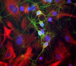
- Immunocytochemistry analysis of SPTAN1 in neurons and glia cells showing specific labeling of neuronal perikarya, axons, and dendrites with alpha-II Spectrin polyclonal antibody (Product # PA5-35383) with a dilution of 1:1,000 (green). Specific labeling of intermediate filaments in glial and fibroblastic cells with anti-vimentin using a dilution of 1:5,000 (red) and nuclear staining with DAPI (blue).
- Submitted by
- Invitrogen Antibodies (provider)
- Main image
- Experimental details
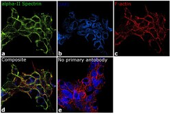
- Immunofluorescence analysis of alpha-II Spectrin was performed using 70% confluent log phase SH-SY5Y cells. The cells were fixed with 4% paraformaldehyde for 10 minutes, permeabilized with 0.1% Triton™ X-100 for 10 minutes, and blocked with 1% BSA for 1 hour at room temperature. The cells were labeled with alpha-II Spectrin Polyclonal Antibody (Product # PA5-35383) at 1:250 dilution in 0.1% BSA and incubated overnight at 4 degree and then labeled with Goat anti-Rabbit IgG (H+L) Superclonal™ Secondary Antibody, Alexa Fluor® 488 conjugate (Product # A27034) at a dilution of 1:2000 for 45 minutes at room temperature (Panel a: green). Nuclei (Panel b: blue) were stained with ProLong™ Diamond Antifade Mountant with DAPI (Product # P36962). F-actin (Panel c: red) was stained with Rhodamine Phalloidin (Product # R415, 1:300). Panel d represents the merged image showing cytoskeletal localization. Panel e represents control cells with no primary antibody to assess background. The images were captured at 60X magnification.
 Explore
 Validate
 Learn
Learn Western blot
 Immunohistochemistry