 Explore
 Validate
 Learn
Learn Western blot
 Immunoprecipitation
Antibody data
- Antibody Data
- Antigen structure
- References [0]
- Comments [0]
- Validations
- Western blot [2]
- Flow cytometry [1]
- Other assay [1]
Submit
Validation data
Reference
Comment
Report error
- Product number
- PA5-16621 - Provider product page

- Provider
- Invitrogen Antibodies
- Product name
- Amphiregulin Polyclonal Antibody
- Antibody type
- Polyclonal
- Antigen
- Synthetic peptide
- Description
- PA5-16621 targets Amphiregulin in IP and WB applications and shows reactivity with Human and mouse samples. The PA5-16621 immunogen is a synthetic 19-mer peptide, corresponding to aa 26-44 from the secreted amphiregulin of human origin.
- Reactivity
- Human, Mouse
- Host
- Rabbit
- Isotype
- IgG
- Vial size
- 500 µL
- Concentration
- 1 mg/mL
- Storage
- 4° C
No comments: Submit comment
Supportive validation
- Submitted by
- Invitrogen Antibodies (provider)
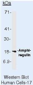
- Main image

- Experimental details
- Western blot of Amphiregulin using Amphiregulin Polyclonal Antibody (Product # PA5-16621) on LS174T Cells.
- Submitted by
- Invitrogen Antibodies (provider)
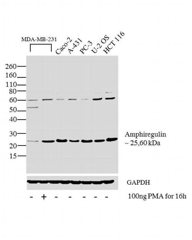
- Main image

- Experimental details
- Western blot analysis was performed on membrane enriched extracts (30 µg lysate) of MDA-MB-231 (Lane 1), MDA-MB-231 treated with PMA (100 ng PMA for 16h) (Lane 2), Caco-2 (Lane 3), A-431 (Lane 4), PC-3 (Lane 5), U-2 OS (Lane 6) and HCT-116 (Lane7).The blots were probed with Amphiregulin Rabbit polyclonal Antibody (Product # PA5-16621, 2 µg/mL) and detected by chemiluminescence using Goat anti-Rabbit IgG (H+L) Superclonal™ Secondary Antibody, HRP conjugate (Product # A27036, 0.4 µg/mL, 1:2500 dilution). A 25 kDa band corresponding to Amphiregulin was observed across the cell lines tested and was enhanced upon treatment in MDA-MB-231 cell line. Apart from desired band of 25 KDa, ~60 kDa band is seen across the cell lines showing the glycosylated form of the protein. Known quantity of protein samples were electrophoresed using Novex® NuPAGE® 4-12 % Bis-Tris gel (Product # NP0322BOX), XCell SureLock™ Electrophoresis System (Product # EI0002) and Novex® Sharp Pre-Stained Protein Standard (Product # LC5800). Resolved proteins were then transferred onto a nitrocellulose membrane with iBlot® 2 Dry Blotting System (Product # IB21001). The membrane was probed with the relevant primary and secondary Antibody following blocking with 5% skimmed milk. Chemiluminescent detection was performed using Pierce™ ECL Western Blotting Substrate (Product # 32106).
Supportive validation
- Submitted by
- Invitrogen Antibodies (provider)
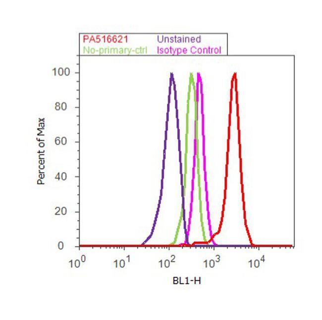
- Main image
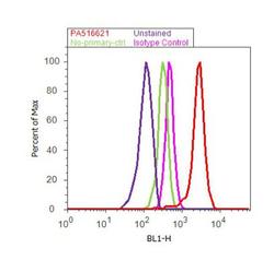
- Experimental details
- Flow cytometry analysis of Amphiregulin was done on MDA-MB-231 cells. Cells were fixed with 70% ethanol for 10 minutes, permeabilized with 0.25% Triton™ X-100 for 20 minutes, and blocked with 5% BSA for 30 minutes at room temperature. Cells were labeled with Amphiregulin Rabbit Polyclonal Antibody (PA5-16621, red histogram) or with rabbit isotype control (pink histogram) at 3-5 µg/million cells in 2.5% BSA. After incubation at room temperature for 2 hours, the cells were labeled with Alexa Fluor® 488 Goat Anti-Rabbit Secondary Antibody (A11008) at a dilution of 1:400 for 30 minutes at room temperature. The representative 10,000 cells were acquired and analyzed for each sample using an Attune® Acoustic Focusing Cytometer. The purple histogram represents unstained control cells and the green histogram represents no-primary-antibody control..
Supportive validation
- Submitted by
- Invitrogen Antibodies (provider)
- Main image
- Experimental details
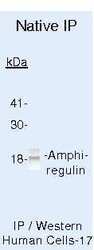
- Immunoprecipitation of Amphiregulin using Amphiregulin Polyclonal Antibody (Product # PA5-16621) on Native Human LS174T Cells.
