Explore
 Validate
 Learn
Learn Western blot
 Immunohistochemistry
Antibody data
- Antibody Data
- Antigen structure
- References [0]
- Comments [0]
- Validations
- Western blot [3]
- Immunohistochemistry [4]
Submit
Validation data
Reference
Comment
Report error
- Product number
- HPA025826 - Provider product page

- Provider
- Atlas Antibodies
- Proper citation
- Atlas Antibodies Cat#HPA025826, RRID:AB_1854106
- Product name
- Anti-MRPL37
- Antibody type
- Polyclonal
- Reactivity
- Human
- Host
- Rabbit
- Conjugate
- Unconjugated
- Antigen sequence
SATWNRESLLLQVRGSGGARLSTKDPLPTIASREE
IEATKNHVLETFYPISPIIDLHECNIYDVK- Isotype
- IgG
- Vial size
- 100 µl
- Storage
- Store at +4°C for short term storage. Long time storage is recommended at -20°C.
No comments: Submit comment
Supportive validation
Supportive validation
- Submitted by
- Atlas Antibodies (provider)
- Enhanced method
- Genetic validation
- Main image

- Experimental details
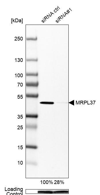
- Western blot analysis in Hep-G2 cells transfected with control siRNA, target specific siRNA probe #1, using Anti-MRPL37 antibody. Remaining relative intensity is presented. Loading control: Anti-PPIB.
- Submitted by
- Atlas Antibodies (provider)
- Enhanced method
- Independent antibody validation
- Main image
- Experimental details
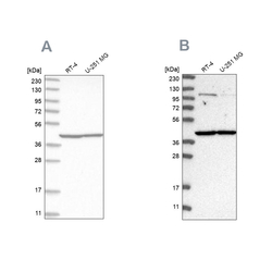
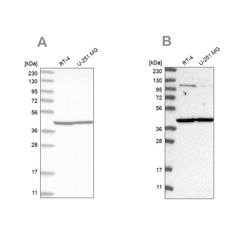
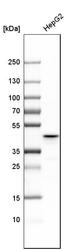
- Western blot analysis using Anti-MRPL37 antibody HPA025826 (A) shows similar pattern to independent antibody HPA025767 (B).
Supportive validation
- Submitted by
- Atlas Antibodies (provider)
- Main image
- Experimental details
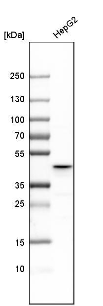
- Western blot analysis in human cell line HepG2.
- Sample type
- HUMAN
Enhanced validation
Supportive validation
- Submitted by
- Atlas Antibodies (provider)
- Enhanced method
- Orthogonal validation
- Main image

- Experimental details
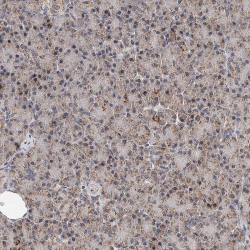
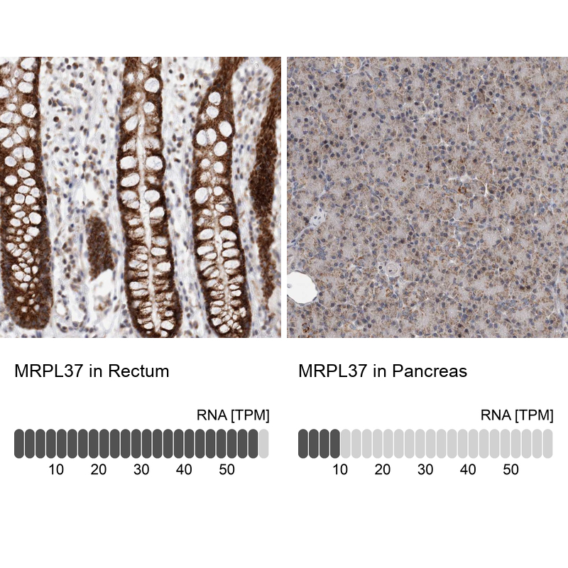
- Immunohistochemistry analysis in human rectum and pancreas tissues using Anti-MRPL37 antibody. Corresponding MRPL37 RNA-seq data are presented for the same tissues.
- Sample type
- HUMAN
Supportive validation
- Submitted by
- Atlas Antibodies (provider)
- Main image
- Experimental details
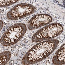
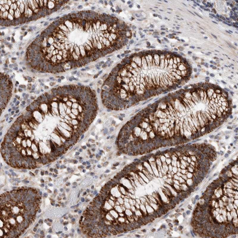
- Immunohistochemical staining of human colon shows strong cytoplasmic positivity in glandular cells.
- Submitted by
- Atlas Antibodies (provider)
- Main image

- Experimental details
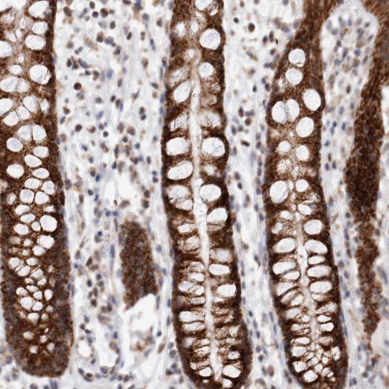
- Immunohistochemical staining of human rectum shows high expression.
- Sample type
- HUMAN
- Submitted by
- Atlas Antibodies (provider)
- Main image
- Experimental details
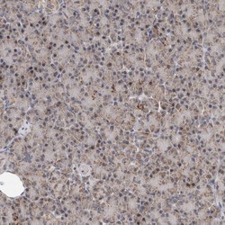
- Immunohistochemical staining of human pancreas shows low expression as expected.
- Sample type
- HUMAN