 Explore
 Validate
 Learn
Learn Western blot
Antibody data
- Antibody Data
- Antigen structure
- References [4]
- Comments [0]
- Validations
- Western blot [3]
- Immunocytochemistry [1]
- Immunoprecipitation [1]
Submit
Validation data
Reference
Comment
Report error
- Product number
- GTX108356 - Provider product page

- Provider
- GeneTex
- Proper citation
- GeneTex Cat#GTX108356, RRID:AB_1950487
- Product name
- HES1 antibody [N1], N-term
- Antibody type
- Polyclonal
- Reactivity
- Human, Mouse, Rat
- Host
- Rabbit
Submitted references Salvianolic Acid B Enhances Hepatic Differentiation of Human Embryonic Stem Cells Through Upregulation of WNT Pathway and Inhibition of Notch Pathway.
Inflammation-induced miRNA-155 inhibits self-renewal of neural stem cells via suppression of CCAAT/enhancer binding protein β (C/EBPβ) expression.
Notch-1 signaling promotes the malignant features of human breast cancer through NF-κB activation.
Hes-1 SUMOylation by protein inhibitor of activated STAT1 enhances the suppressing effect of Hes-1 on GADD45α expression to increase cell survival.
Chen J, Tschudy-Seney B, Ma X, Zern MA, Liu P, Duan Y
Stem cells and development 2018 Feb 15;27(4):252-261
Stem cells and development 2018 Feb 15;27(4):252-261
Inflammation-induced miRNA-155 inhibits self-renewal of neural stem cells via suppression of CCAAT/enhancer binding protein β (C/EBPβ) expression.
Obora K, Onodera Y, Takehara T, Frampton J, Hasei J, Ozaki T, Teramura T, Fukuda K
Scientific reports 2017 Feb 27;7:43604
Scientific reports 2017 Feb 27;7:43604
Notch-1 signaling promotes the malignant features of human breast cancer through NF-κB activation.
Li L, Zhao F, Lu J, Li T, Yang H, Wu C, Liu Y
PloS one 2014;9(4):e95912
PloS one 2014;9(4):e95912
Hes-1 SUMOylation by protein inhibitor of activated STAT1 enhances the suppressing effect of Hes-1 on GADD45α expression to increase cell survival.
Chiou HY, Liu SY, Lin CH, Lee EH
Journal of biomedical science 2014 Jun 4;21:53
Journal of biomedical science 2014 Jun 4;21:53
No comments: Submit comment
Supportive validation
- Submitted by
- GeneTex (provider)
- Main image
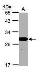
- Experimental details
- Sample(30 ?g whole cell lysate)A:A431(GTX27909) 12% SDS PAGEGTX108356 diluted at 1:1000The HRP-conjugated anti-rabbit IgG antibody (GTX213110-01) was used to detect the primary antibody.

- Submitted by
- GeneTex (provider)
- Main image
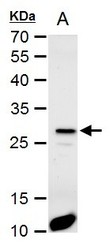
- Experimental details
- HES1 antibody [N1], N-term detects HES1 protein by western blot analysis.A.50 ?g mouse kidney extract12% SDS-PAGEHES1 antibody [N1], N-term (GTX108356) dilution: 1:1000 The HRP-conjugated anti-rabbit IgG antibody (GTX213110-01) was used to detect the primary antibody.

- Submitted by
- GeneTex (provider)
- Main image

- Experimental details
- HES1 antibody [N1], N-term detects HES1 protein by western blot analysis.A. 30 ?g PC-12 whole cell extract12% SDS-PAGEHES1 antibody [N1], N-term (GTX108356) dilution: 1:1000 The HRP-conjugated anti-rabbit IgG antibody (GTX213110-01) was used to detect the primary antibody.

Supportive validation
- Submitted by
- GeneTex (provider)
- Main image

- Experimental details
- HES1 antibody [N1], N-term detects HES1 protein at nucleus by immunofluorescent analysis.Sample: HeLa cells were fixed in 4% paraformaldehyde at RT for 15 min.Green: HES1 protein stained by HES1 antibody [N1], N-term (GTX108356) diluted at 1:2000.Red: alpha Tubulin, a cytoskeleton marker, stained by alpha Tubulin antibody [GT114] (GTX628802) diluted at 1:1000.Blue: Hoechst 33342 staining.

Supportive validation
- Submitted by
- GeneTex (provider)
- Main image
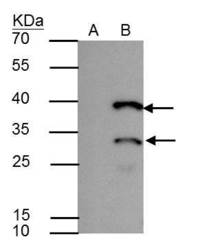
- Experimental details
- HES1 antibody immunoprecipitates HES1 protein in IP experiments. IP Sample: A431 whole cell lysate/extract A : Control with 2.5 £gg of pre-immune rabbit IgG B : Immunoprecipitation of HES1 by 2.5 £gg of HES1 antibody (GTX108356) 12% SDS-PAGE The immunoprecipitated HES1 protein was detected by HES1 antibody (GTX108356) diluted at 1 : 1000. EasyBlot anti-rabbit IgG (HRP) (GTX221666-01) was used as a secondary reagent.
