 Explore
 Validate
 Learn
Learn Western blot
Antibody data
- Antibody Data
- Antigen structure
- References [0]
- Comments [0]
- Validations
- Western blot [2]
- Immunocytochemistry [1]
- Immunohistochemistry [5]
- Flow cytometry [1]
Submit
Validation data
Reference
Comment
Report error
- Product number
- 20887-1-AP - Provider product page

- Provider
- Invitrogen Antibodies
- Product name
- Caldesmon Polyclonal Antibody
- Antibody type
- Polyclonal
- Antigen
- Other
- Description
- Immunogen sequence: EKPKRGSIG ENQIKDEKIK KDKEPKEEVK SFMDRKKGFT EVKSQNGEFM THKLKHTENT FSRPGGRASV DTKEAEGAPQ VEAGKRLEEL RRRRGETESE EFEKLKQKQQ EAALELEELK KKREERRKVL EEEEQRRKQE EADRKLREEE EKRRLKEEIE RRRAEAAEKR QKMPEDGLSD DKKPFKCFTP KGSSLKIEER AEFLNKSVQK SSGVKSTHQA AIVSKIDSRL EQYTSAIEGT KSAKPTKPAA SDLPVPAEGV RNIKSMWEKG NVFSSPTAAG TPNKETAGLK VGVSSRINEW LTKTPDGNKS PAPKPSDLRP GDVSSKRNLW EKQSVDKVTS PTKV (196-538 aa encoded by B C040354)
- Reactivity
- Human, Mouse
- Host
- Rabbit
- Isotype
- IgG
- Vial size
- 150 µL
- Concentration
- 0.13 mg/mL
- Storage
- -20°C
No comments: Submit comment
Supportive validation
- Submitted by
- Invitrogen Antibodies (provider)
- Main image

- Experimental details
- Mouse small intestine tissue were subjected to SDS PAGE followed by western blot with 20887-1-AP (Caldesmon antibody) at dilution of 1:500 incubated at room temperature for 1.5 hours.

- Submitted by
- Invitrogen Antibodies (provider)
- Main image

- Experimental details
- PC-3 cells were subjected to SDS PAGE followed by western blot with 20887-1-AP (Caldesmon antibody) at dilution of 1:500 incubated at room temperature for 1.5 hours.

Supportive validation
- Submitted by
- Invitrogen Antibodies (provider)
- Main image
- Experimental details
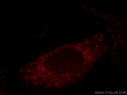
- Immunofluorescent analysis of HepG2 cells, using CALD1 antibody 20887-1-AP at 1:25 dilution and Rhodamine-labeled goat anti-rabbit IGG (red).

Supportive validation
- Submitted by
- Invitrogen Antibodies (provider)
- Main image

- Experimental details
- Immunofluorescent analysis of ( 4% PFA ) fixed mouse heart tissue using 20887-1-AP (Caldesmon antibody) at dilution of 1:50 and Alexa Fluor 488-conjugated AffiniPure Goat Anti-Rabbit IGG (H+L).

- Submitted by
- Invitrogen Antibodies (provider)
- Main image

- Experimental details
- Immunohistochemistry of paraffin-embedded human colon tissue slide using 20887-1-AP (Caldesmon antibody at dilution of 1:50 (under 10x lens).

- Submitted by
- Invitrogen Antibodies (provider)
- Main image

- Experimental details
- Immunohistochemistry of paraffin-embedded human colon tissue slide using 20887-1-AP (Caldesmon antibody at dilution of 1:50 (under 40x lens).

- Submitted by
- Invitrogen Antibodies (provider)
- Main image

- Experimental details
- Immunohistochemistry of paraffin-embedded human colon cancer tissue slide using 20887-1-AP ( Caldesmon antibody at dilution of 1:100 (under 10x lens). heat mediated antigen retrieved with Tris-EDTA buffer (pH 9).

- Submitted by
- Invitrogen Antibodies (provider)
- Main image

- Experimental details
- Immunohistochemistry of paraffin-embedded human colon cancer tissue slide using 20887-1-AP ( Caldesmon antibody at dilution of 1:100 (under 40x lens). heat mediated antigen retrieved with Tris-EDTA buffer (pH 9).

Supportive validation
- Submitted by
- Invitrogen Antibodies (provider)
- Main image
- Experimental details
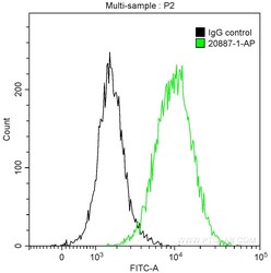
- 1X10^6 HeLa cells were intracellularly stained with 0.2 µg Anti-Human Caldesmon (Product # 20887-1-AP) and CoraLite®488-Conjugated AffiniPure Goat Anti-Rabbit IgG(H+L) at dilution 1:1,000 (green), and 0.2 µg Control Antibody. Cells were fixed with 90% MeOH.
