 Explore
 Validate
 Learn
Learn Western blot
 Immunocytochemistry
Antibody data
- Antibody Data
- Antigen structure
- References [3]
- Comments [0]
- Validations
- Western blot [2]
- Immunocytochemistry [1]
- Immunohistochemistry [6]
Submit
Validation data
Reference
Comment
Report error
- Product number
- HPA017330 - Provider product page

- Provider
- Atlas Antibodies
- Proper citation
- Atlas Antibodies Cat#HPA017330, RRID:AB_1845917
- Product name
- Anti-CALD1
- Antibody type
- Polyclonal
- Reactivity
- Human
- Host
- Rabbit
- Conjugate
- Unconjugated
- Antigen sequence
MTHKLKHTENTFSRPGGRASVDTKEAEGAPQVEAG
KRLEELRRRRGETESEEFEKLKQKQQEAALELEEL
KKKREERRKVLEEEEQRRKQEEADRKLREEEEKRR
LKEEIERRRAEAAEKRQKMPEDGLSDD- Isotype
- IgG
- Vial size
- 100 µl
- Storage
- Store at +4°C for short term storage. Long time storage is recommended at -20°C.
Submitted references Antibodies Biotinylated Using a Synthetic Z-domain from Protein A Provide Stringent In Situ Protein Detection
Profiling post-centrifugation delay of serum and plasma with antibody bead arrays
Histochemical localization of caldesmon in the CNS and ganglia of the mouse.
Andersson S, Konrad A, Ashok N, Ponten F, Hober S, Asplund A
Journal of Histochemistry & Cytochemistry 2013 October;61(11):773-784
Journal of Histochemistry & Cytochemistry 2013 October;61(11):773-784
Profiling post-centrifugation delay of serum and plasma with antibody bead arrays
Qundos U, Hong M, Tybring G, Divers M, Odeberg J, Uhlen M, Nilsson P, Schwenk J
Journal of Proteomics 2013 December;95
Journal of Proteomics 2013 December;95
Histochemical localization of caldesmon in the CNS and ganglia of the mouse.
Köhler CN
The journal of histochemistry and cytochemistry : official journal of the Histochemistry Society 2011 May;59(5):504-17
The journal of histochemistry and cytochemistry : official journal of the Histochemistry Society 2011 May;59(5):504-17
No comments: Submit comment
Enhanced validation
Enhanced validation
- Submitted by
- Atlas Antibodies (provider)
- Enhanced method
- Orthogonal validation
- Main image

- Experimental details
- Western blot analysis in human cell lines U-251MG and MCF-7 using Anti-CALD1 antibody. Corresponding CALD1 RNA-seq data are presented for the same cell lines. Loading control: Anti-GAPDH.

Enhanced validation
- Submitted by
- Atlas Antibodies (provider)
- Enhanced method
- Independent antibody validation
- Main image

- Experimental details
- Western blot analysis using Anti-CALD1 antibody HPA017330 (A) shows similar pattern to independent antibody HPA008066 (B).

Supportive validation
- Submitted by
- Atlas Antibodies (provider)
- Main image

- Experimental details
- Immunofluorescent staining of human cell line U-2 OS shows localization to plasma membrane & actin filaments.
- Sample type
- HUMAN

Enhanced validation
Enhanced validation
Supportive validation
- Submitted by
- Atlas Antibodies (provider)
- Enhanced method
- Orthogonal validation
- Main image

- Experimental details
- Immunohistochemistry analysis in human smooth muscle and skeletal muscle tissues using Anti-CALD1 antibody. Corresponding CALD1 RNA-seq data are presented for the same tissues.
- Sample type
- HUMAN

Enhanced validation
- Submitted by
- Atlas Antibodies (provider)
- Enhanced method
- Independent antibody validation
- Main image

- Experimental details
- Immunohistochemical staining of human cerebral cortex, placenta, skeletal muscle and smooth muscle using Anti-CALD1 antibody HPA017330 (A) shows similar protein distribution across tissues to independent antibody HPA008066 (B).

Supportive validation
- Submitted by
- Atlas Antibodies (provider)
- Main image
- Experimental details
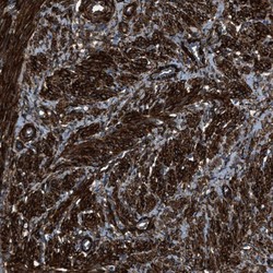
- Immunohistochemical staining of human smooth muscle shows high expression.
- Sample type
- HUMAN

- Submitted by
- Atlas Antibodies (provider)
- Main image

- Experimental details
- Immunohistochemical staining of human skeletal muscle shows low expression as expected.
- Sample type
- HUMAN

- Submitted by
- Atlas Antibodies (provider)
- Main image

- Experimental details
- Immunohistochemical staining of human cerebral cortex using Anti-CALD1 antibody HPA017330.
- Sample type
- HUMAN

- Submitted by
- Atlas Antibodies (provider)
- Main image

- Experimental details
- Immunohistochemical staining of human placenta using Anti-CALD1 antibody HPA017330.
- Sample type
- HUMAN
