 Explore
 Validate
 Learn
Learn Immunocytochemistry
 Immunohistochemistry
Antibody data
- Antibody Data
- Antigen structure
- References [1]
- Comments [0]
- Validations
- Immunocytochemistry [1]
- Immunohistochemistry [7]
Submit
Validation data
Reference
Comment
Report error
- Product number
- HPA021195 - Provider product page

- Provider
- Atlas Antibodies
- Proper citation
- Atlas Antibodies Cat#HPA021195, RRID:AB_1844528
- Product name
- Anti-ACOX1
- Antibody type
- Polyclonal
- Reactivity
- Human
- Host
- Rabbit
- Conjugate
- Unconjugated
- Antigen sequence
AVRHQSEIKPGEPEPQILDFQTQQYKLFPLLATAY
AFQFVGAYMKETYHRINEGIGQGDLSELPELHALT
AGLKAFTSWTANTGIEACRMACGGHGY- Isotype
- IgG
- Vial size
- 100 µl
- Storage
- Store at +4°C for short term storage. Long time storage is recommended at -20°C.
Submitted references Immunofluorescence and fluorescent-protein tagging show high correlation for protein localization in mammalian cells
Stadler C, Rexhepaj E, Singan V, Murphy R, Pepperkok R, Uhlén M, Simpson J, Lundberg E
Nature Methods 2013 February;10(4):315-323
Nature Methods 2013 February;10(4):315-323
No comments: Submit comment
Supportive validation
- Submitted by
- Atlas Antibodies (provider)
- Main image
- Experimental details
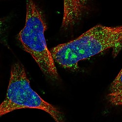
- Immunofluorescent staining of human cell line U-2 OS shows localization to nucleoli & vesicles.
- Sample type
- HUMAN

Enhanced validation
Supportive validation
- Submitted by
- Atlas Antibodies (provider)
- Enhanced method
- Independent antibody validation
- Main image
- Experimental details
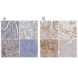
- Immunohistochemical staining of human gastrointestinal, kidney, liver and pancreas using Anti-ACOX1 antibody HPA021195 (A) shows similar protein distribution across tissues to independent antibody HPA028759 (B).

Supportive validation
- Submitted by
- Atlas Antibodies (provider)
- Main image

- Experimental details
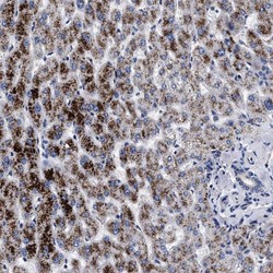
- Immunohistochemical staining of human stomach shows strong positivity in superficial glandular cells.

- Submitted by
- Atlas Antibodies (provider)
- Main image
- Experimental details
- Immunohistochemical staining of human liver shows strong cytoplasmic positivity in hepatocytes.
- Sample type
- HUMAN

- Submitted by
- Atlas Antibodies (provider)
- Main image
- Experimental details
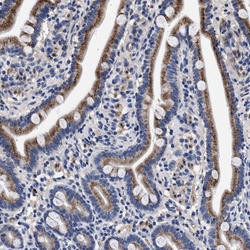
- Immunohistochemical staining of human duodenum shows strong granular cytoplasmic positivity in glandular cells.
- Sample type
- HUMAN

- Submitted by
- Atlas Antibodies (provider)
- Main image
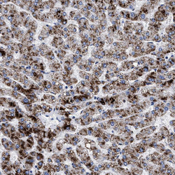
- Experimental details
- Immunohistochemical staining of human liver shows strong granular cytoplasmic positivity in hepatocytes.
- Sample type
- HUMAN

- Submitted by
- Atlas Antibodies (provider)
- Main image

- Experimental details
- Immunohistochemical staining of human kidney shows moderate granular cytoplasmic positivity in cells in tubules.
- Sample type
- HUMAN

- Submitted by
- Atlas Antibodies (provider)
- Main image

- Experimental details
- Immunohistochemical staining of human pancreas shows no positivity in exocrine glandular cells.
- Sample type
- HUMAN
