 Explore
 Validate
 Learn
Learn701685
antibody from Invitrogen Antibodies
Targeting: ATG16L1
APG16L, ATG16A, ATG16L, FLJ10035, WDR30
 Western blot
Antibody data
- Antibody Data
- Antigen structure
- References [0]
- Comments [0]
- Validations
- Western blot [3]
- Immunocytochemistry [1]
- Flow cytometry [1]
Submit
Validation data
Reference
Comment
Report error
- Product number
- 701685 - Provider product page

- Provider
- Invitrogen Antibodies
- Product name
- ATG16L1 Recombinant Rabbit Monoclonal Antibody (17H32L7)
- Antibody type
- Monoclonal
- Antigen
- Other
- Description
- This antibody is predicted to react with Monkey, Rat, Mouse and Rabbit.
- Antibody clone number
- 17H32L7
- Concentration
- 0.5 mg/mL
No comments: Submit comment
Supportive validation
- Submitted by
- Invitrogen Antibodies (provider)
- Main image
- Experimental details
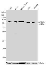
- Western blot analysis was performed on whole cell extracts (30 µg lysate) of Hela (Lane1), PC-3 (Lane 2), Mouse testis (Lane 3), HEL (Lane 4) and U-87MG (Lane 5). The blots were probed with Anti-ATG16L Recombinant Rabbit Monoclonal Antibody (Product # 701685, 4-5 µg/mL) and detected by chemiluminescence using Goat anti-Rabbit IgG (H+L) Superclonal™ Secondary Antibody, HRP conjugate (Product # A27036, 0.4 µg/mL, 1:2500 dilution). A 65 kDa band corresponding to ATG16L was observed across cell lines tested. Known quantity of protein samples were electrophoresed using Novex® NuPAGE® 4-12% Bis-Tris gel (Product # NP0321BOX), XCell SureLock™ Electrophoresis System (Product # EI0002) and Novex® Sharp Pre-Stained Protein Standard (Product # LC5800). Resolved proteins were then transferred onto a nitrocellulose membrane with iBlot® Dry Blotting System (Product # IB21001). The membrane was probed with the relevant primary and secondary Antibody following blocking with 5% skimmed milk. Chemiluminescent detection was performed using Pierce™ ECL Western blotting Substrate (Product # 32106).

- Submitted by
- Invitrogen Antibodies (provider)
- Main image

- Experimental details
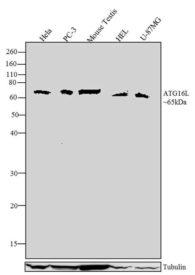
- Western blot analysis was performed on whole cell extracts (30 µg lysate) of Hela (Lane1), PC-3 (Lane 2), Mouse testis (Lane 3), HEL (Lane 4) and U-87MG (Lane 5). The blots were probed with Anti-ATG16L Recombinant Rabbit Monoclonal Antibody (Product # 701685, 4-5 µg/mL) and detected by chemiluminescence using Goat anti-Rabbit IgG (H+L) Superclonal™ Secondary Antibody, HRP conjugate (Product # A27036, 0.4 µg/mL, 1:2500 dilution). A 65 kDa band corresponding to ATG16L was observed across cell lines tested. Known quantity of protein samples were electrophoresed using Novex® NuPAGE® 4-12% Bis-Tris gel (Product # NP0321BOX), XCell SureLock™ Electrophoresis System (Product # EI0002) and Novex® Sharp Pre-Stained Protein Standard (Product # LC5800). Resolved proteins were then transferred onto a nitrocellulose membrane with iBlot® Dry Blotting System (Product # IB21001). The membrane was probed with the relevant primary and secondary Antibody following blocking with 5% skimmed milk. Chemiluminescent detection was performed using Pierce™ ECL Western blotting Substrate (Product # 32106).
- Submitted by
- Invitrogen Antibodies (provider)
- Main image
- Experimental details
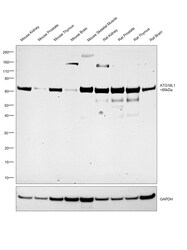
- Western blot was performed using Anti-ATG16L1 Recombinant Rabbit Monoclonal Antibody (17H32L7) (Product # 701685) and a 65 kDa band corresponding to ATG16L1 was observed across all tissues tested. Whole cell extracts (30 µg lysate) of Mouse Kidney (Lane 1), Mouse Prostate (Lane 2), Mouse Thymus (Lane 3), Mouse Brain (Lane 4), Mouse Skeletal Muscle (Lane 5), Rat Kidney (Lane 6), Rat Prostate (Lane 7), Rat Thymus (Lane 8), Rat Brain (Lane 9) were electrophoresed using NuPAGE™ 4-12% Bis-Tris Protein Gel (Product # NP0321BOX). Resolved proteins were then transferred onto a Nitrocellulose membrane (Product # IB23001) by iBlot® 2 Dry Blotting System (Product # IB21001). The blot was probed with the primary antibody (1 µg/mL dilution) and detected by chemiluminescence with Goat anti-Rabbit IgG (H+L) Superclonal™ Recombinant Secondary Antibody, HRP (Product # A27036, 1:4000 dilution) using the iBright FL 1000 (Product # A32752). Chemiluminescent detection was performed using Novex® ECL Reagent Kit (Product # WP20005).

Supportive validation
- Submitted by
- Invitrogen Antibodies (provider)
- Main image
- Experimental details
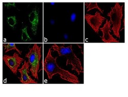
- Immunofluorescence was performed on fixed and permeabilized HeLa cells for detection of ATG16L using Anti-ATG16L Recombinant Rabbit Monoclonal Antibody (Product # 701685, 2 µg/mL) and labeled with Goat anti-Rabbit IgG (H+L) Superclonal™ Secondary Antibody, Alexa Fluor® 488 conjugate (Product # A27034, 1:2000). Panel a) shows representative cells that were stained for detection and localization of ATG16L protein (green), Panel b) is stained for nuclei (blue) using SlowFade® Gold Antifade Mountant with DAPI (Product # S36938). Panel c) represents cytoskeletal F-actin staining using Alexa Fluor® 555 Rhodamine Phalloidin (Product # R415, 1:300). Panel d) is a composite image of Panels a, b and c clearly demonstrating cytoplasmic localization of ATG16L Panel e) represents control cells with no primary Antibody to assess background.

Supportive validation
- Submitted by
- Invitrogen Antibodies (provider)
- Main image

- Experimental details
- Flow Cytometry analysis of ATG16L was performed on HeLa cells labeled with ABfinity™ Anti-ATG16L Recombinant Rabbit Monoclonal Antibody (Product# 701685, 2-4 ug/ 1M cells) or with Rabbit isotype control and detected with Goat anti-Rabbit IgG (H+L) Superclonal™ Secondary Antibody, (Alexa Fluor® 488 conjugate, Product # A27034, 0.4 ug/ml, 1:2500) as represented by the red and yellow histograms respectively. The purple histogram represents unstained control cells and the green histogram represents no-primary-Antibody control. A representative of 10,000 cells were acquired and analyzed for each sample using an Attune® Acoustic Focusing Cytometer (4468770).
