 Explore
 Validate
 Learn
LearnAP32161PU-N
antibody from Acris Antibodies GmbH
Targeting: MAP1LC3A
ATG8E, LC3, LC3A, MAP1ALC3, MAP1BLC3
 Western blot
Antibody data
- Antibody Data
- Antigen structure
- References [0]
- Comments [0]
- Validations
- Western blot [2]
- Immunocytochemistry [2]
- Immunohistochemistry [1]
Submit
Validation data
Reference
Comment
Report error
- Product number
- AP32161PU-N - Provider product page

- Provider
- Acris Antibodies GmbH
- Product name
- anti LC3 (N-term)
- Antibody type
- Polyclonal
- Antigen
- KLH conjugated synthetic peptide between 1~30 amino acids from the N-term of Human LC3 (APG8a).
- Reactivity
- Human, Mouse, Rat
- Host
- Rabbit
- Vial size
- 0.4 ml
- Concentration
- lot specific
No comments: Submit comment
Supportive validation
- Submitted by
- Acris Antibodies GmbH (provider)
- Main image

- Experimental details
- Western blot analysis using LC3 Antibody Cat.-No AP32161PU-N in Mouse brain tissue lysates (35ug/lane).

- Submitted by
- Acris Antibodies GmbH (provider)
- Main image

- Experimental details
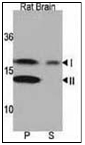
- Western blot analysis of LC3 Antibody Cat.-No AP32161PU-N in Rat brain lysate. Both lipidated (arrow, II) and non-lipidated APG8a (arrow, I) were detected in membrane fraction (P) but only non-lipidated LC3 was detected in soluble fraction (S).
Supportive validation
- Submitted by
- Acris Antibodies GmbH (provider)
- Main image

- Experimental details
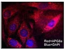
- Immunofluorescent staining of Mouse brain cells using LC3 Antibody Cat.-No AP32161PU-N
- Submitted by
- Acris Antibodies GmbH (provider)
- Main image

- Experimental details
- LC3 and the CLN3-encoded protein, battenin, co-immunostaining. Wild-type and mutant cerebellar cells were costained with anti-LC3 Antibody Cat.-No AP32161PU-NÂ (Red) and a monoclonal anti-Battenin Antibody(Green) and images were captured by Confocal Microscopy. Note the numerous larger vesicular structures, typical of mature AVs, stained with both the LC3 and battenin antibodies in wild-type cells (arrows), which are rare in mutant cells. Blue=DAPI. Protocol and data courtesy of Dr. Susan Cotman, Center for Human Genetic Research, Massachusetts General Hospital.

Supportive validation
- Submitted by
- Acris Antibodies GmbH (provider)
- Main image
- Experimental details
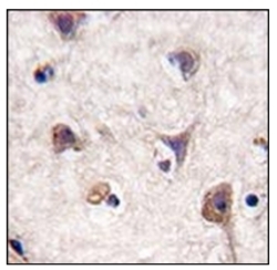
- Formalin-fixed and paraffin-embedded human brain tissue reacted with Autophagy LC3 Antibody Cat.-No AP32161PU-N, which was peroxidase-conjugated to the secondary antibody, followed by DAB staining. This data demonstrates the use of this antibody for immunohistochemistry; clinical relevance has not been evaluated.
