AM32460PU-N
antibody from Acris Antibodies GmbH
Targeting: MAP1LC3A
ATG8E, LC3, LC3A, MAP1ALC3, MAP1BLC3
Antibody data
- Antibody Data
- Antigen structure
- References [0]
- Comments [0]
- Validations
- Western blot [2]
- Immunocytochemistry [2]
- Immunohistochemistry [2]
Submit
Validation data
Reference
Comment
Report error
- Product number
- AM32460PU-N - Provider product page

- Provider
- Acris Antibodies GmbH
- Product name
- anti LC3
- Antibody type
- Monoclonal
- Antigen
- Full length recombinant protein of Human LC3 (APG8).
- Reactivity
- Human, Mouse, Rat
- Host
- Mouse
- Isotype
- IgG
- Antibody clone number
- 166AT1234
- Vial size
- 0.4 ml
- Concentration
- lot specific
No comments: Submit comment
Supportive validation
- Submitted by
- Acris Antibodies GmbH (provider)
- Main image

- Experimental details
- Western blot analysis of LC3 Antibody Cat.-No AM32460PU-N (Clone 166AT1234) at 8 µg/ml. Lane 1: Y79 (soluble fraction of cell extract). Lane 2: 293 transfected with human LC3 (whole cell extract).
- Submitted by
- Acris Antibodies GmbH (provider)
- Main image

- Experimental details
- Western blot analysis of LC3 Antibody Cat.-No AM32460PU-N (Clone 166AT1234) in Hela cell lysates, which were treated with rapamycin or bafilomycin overnight. Data courtesy of Dr. David Rubinsztein, Cambridge Institute for Medical Research.
Supportive validation
- Submitted by
- Acris Antibodies GmbH (provider)
- Main image

- Experimental details
- Mouse cerebellar cell lines stably expressing human GFP-LC3 fusion protein were evaluated by Immunocytochemistry using the LC3 Antibody Cat.-No AM32460PU-N (Clone 166AT1234). Punctuate autophagic cell vesicles, with numerous GFP-LC3 vesicles are displayed. Conditions: 1:1 methanol:acetone fixation, 0.2% saponin permeabilization.
- Submitted by
- Acris Antibodies GmbH (provider)
- Main image

- Experimental details
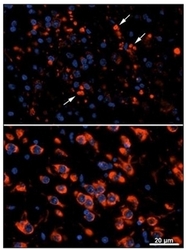
- Cathepsin D WT vs KO (postnatal day 25) mouse thalamus exhibiting clumped immunoreactivity for LC3 in the KO brain (arrows, upper panel) and relative absence of clumped LC3 immunoreactivity in the WT control (staining is diffuse and cytoplasmic, lower panel). Data courtesy of Dr. John Shacka, Neuropathology Division, University of Alabama at Birmingham.
Supportive validation
- Submitted by
- Acris Antibodies GmbH (provider)
- Main image

- Experimental details
- Formalin-fixed and paraffin-embedded human hepatocarcinoma reacted with Autophagy LC3 Antibody Cat.-No AM32460PU-N (Clone 166AT1234), which was peroxidase-conjugated to the secondary antibody, followed by DAB staining. This data demonstrates the use of this antibody for immunohistochemistry. Clinical relevance has not been evaluated.
- Submitted by
- Acris Antibodies GmbH (provider)
- Main image

- Experimental details
- 10X( lower panel) and 20X (upper panel) Immunohistochemistry images from muscle tissue of a diseased mouse off Dox after 5 weeks on regular food. Several fibers that have autophagic vesicles throughout are visible. Primary antibody used is LC3 Antibody Cat.-No AM32460PU-N (Clone 166AT1234). Data courtesy of Dr. Christy Caudill, Cincinnati Children's Hospital Medical Center.
 Explore
 Validate
 Learn
Learn Western blot