 Explore
 Validate
 Learn
Learn Western blot
Antibody data
- Antibody Data
- Antigen structure
- References [1]
- Comments [0]
- Validations
- Western blot [1]
- Immunohistochemistry [2]
- Flow cytometry [1]
- Other assay [1]
Submit
Validation data
Reference
Comment
Report error
- Product number
- 44-1264G - Provider product page

- Provider
- Invitrogen Antibodies
- Product name
- Phospho-SGK1 (Ser422) Polyclonal Antibody
- Antibody type
- Polyclonal
- Antigen
- Synthetic peptide
- Reactivity
- Human
- Host
- Rabbit
- Isotype
- IgG
- Vial size
- 100 µL
- Storage
- -20°C
Submitted references Expression Profiles of GILZ and SGK-1 in Potentially Malignant and Malignant Human Oral Lesions.
Mozaffari MS, Abdelsayed R
Frontiers in oral health 2021;2:675288
Frontiers in oral health 2021;2:675288
No comments: Submit comment
Supportive validation
- Submitted by
- Invitrogen Antibodies (provider)
- Main image

- Experimental details
- Western blot analysis of Phospho-SGK1 (Ser422) using a polyclonal antibody (Product # 44-1264G). Samples (NIH3T3 fibroblast lysates, spiked with 100 ng of active tagged-SGK1 protein [Product # PV3818]) were resolved on a 10% polyacrylamide gel and transferred to PVDF. The membrane was blocked with a 3% BSA-TBST buffer for one hour at room temperature and then incubated with the SGK1 [pS422] antibody for two hours at room temperature in 3% BSA-TBST buffer, following prior incubation with: no peptide (1), the non-phosphopeptide corresponding to the phosphopeptide immunogen (2), a generic phosphoserine-containing peptide (3), or the phosphopeptide immunogen corresponding to SGK1 [pS422] (4). After washing, the membrane was incubated with goat F(ab’)2 anti-rabbit IgG HRP conjugate and signals were detected using the SuperSignal™ reagent.

Supportive validation
- Submitted by
- Invitrogen Antibodies (provider)
- Main image

- Experimental details
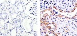
- Immunohistochemistry analysis of SGK1 (PS422) showing staining in the cytoplasm of paraffin-embedded human kidney tissue (right) compared to a negative control without primary antibody (left). To expose target proteins, antigen retrieval was performed using 10mM sodium citrate (pH 6.0), microwaved for 8-15 min. Following antigen retrieval, tissues were blocked in 3% H2O2-methanol for 15 min at room temperature, washed with ddH2O and PBS, and then probed with a SGK1 (PS422) Rabbit Polyclonal Antibody (Product # 44-1264G) diluted in 3% BSA-PBS at a dilution of 1:20 overnight at 4°C in a humidified chamber. Tissues were washed extensively in PBST and detection was performed using an HRP-conjugated secondary antibody followed by colorimetric detection using a DAB kit. Tissues were counterstained with hematoxylin and dehydrated with ethanol and xylene to prep for mounting.

- Submitted by
- Invitrogen Antibodies (provider)
- Main image
- Experimental details
- Immunohistochemistry analysis of SGK1 (PS422) showing staining in the cytoplasm of paraffin-embedded human kidney tissue (right) compared to a negative control without primary antibody (left). To expose target proteins, antigen retrieval was performed using 10mM sodium citrate (pH 6.0), microwaved for 8-15 min. Following antigen retrieval, tissues were blocked in 3% H2O2-methanol for 15 min at room temperature, washed with ddH2O and PBS, and then probed with a SGK1 (PS422) Rabbit Polyclonal Antibody (Product # 44-1264G) diluted in 3% BSA-PBS at a dilution of 1:20 overnight at 4°C in a humidified chamber. Tissues were washed extensively in PBST and detection was performed using an HRP-conjugated secondary antibody followed by colorimetric detection using a DAB kit. Tissues were counterstained with hematoxylin and dehydrated with ethanol and xylene to prep for mounting.

Supportive validation
- Submitted by
- Invitrogen Antibodies (provider)
- Main image
- Experimental details
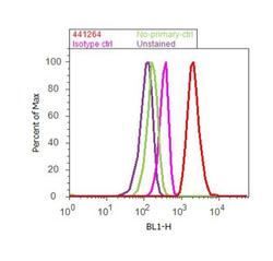
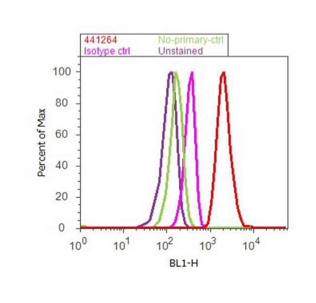
- Flow cytometry analysis of SGK1 [pS422] was done on A549 cells treated with TGF beta (20ng/mL, 15 minutes). Cells were fixed with 70% ethanol for 10 minutes, permeabilized with 0.25% Triton™ X-100 for 20 minutes, and blocked with 5% BSA for 30 minutes at room temperature. Cells were labeled with SGK1 [pS422] Rabbit Polyclonal Antibody (441264G, red histogram) or with rabbit isotype control (pink histogram) at 3-5 ug/million cells in 2.5% BSA. After incubation at room temperature for 2 hours, the cells were labeled with Alexa Fluor® 488 Goat Anti-Rabbit Secondary Antibody (A11008) at a dilution of 1:400 for 30 minutes at room temperature. The representative 10,000 cells were acquired and analyzed for each sample using an Attune® Acoustic Focusing Cytometer. The purple histogram represents unstained control cells and the green histogram represents no-primary-antibody control.
Supportive validation
- Submitted by
- Invitrogen Antibodies (provider)
- Main image

- Experimental details
- Figure 3 (A,B) show SGK-1 and phosphorylated SGK-1 (pSGK-1) immunostaining, respectively, for tissue specimens of the three subjects for each condition (x200). Benign keratosis, mild-moderate dysplasia, and severe dysplasia show prominent positive cytoplasmic staining and variable nuclear staining throughout the full thickness of the epithelium for SGK-1 (A) . It is noteworthy, however, that some cells did not show positive staining. The squamous cell carcinoma cases also manifested prominent cytoplasmic and some nuclear staining throughout the malignant epithelial cells for SGK-1 (A) . (B) shows that benign keratosis, mild-moderate dysplasia, and severe dysplasia display prominent positive cell membrane staining, without nuclear staining, throughout full thickness of the epithelium for pSGK-1. The squamous cell carcinoma cases exhibited positive cell membrane staining throughout malignant epithelial cells for pSGK-1 (B) x200.
