 Explore
 Validate
 Learn
Learn Western blot
 Immunocytochemistry
Antibody data
- Antibody Data
- Antigen structure
- References [0]
- Comments [0]
- Validations
- Western blot [3]
- Immunocytochemistry [1]
- Immunohistochemistry [6]
Submit
Validation data
Reference
Comment
Report error
- Product number
- HPA023034 - Provider product page

- Provider
- Atlas Antibodies
- Proper citation
- Atlas Antibodies Cat#HPA023034, RRID:AB_1855095
- Product name
- Anti-PCYT2
- Antibody type
- Polyclonal
- Reactivity
- Human
- Host
- Rabbit
- Conjugate
- Unconjugated
- Antigen sequence
TAELLSHFKVDLVCHGKTEIIPDRDGSDPYQEPKR
RGIFRQIDSGSNLTTDLIVQRIITNRLEYEARNQK
KEAKELAFLEAARQQAAQPLGERDGDF- Isotype
- IgG
- Vial size
- 100 µl
- Storage
- Store at +4°C for short term storage. Long time storage is recommended at -20°C.
No comments: Submit comment
Enhanced validation
Supportive validation
Supportive validation
- Submitted by
- 57e50c6745390
- Enhanced method
- Orthogonal validation
- Main image

- Experimental details
- Orthogonal validation by targeted mass spectrometry with stable isotope labeled antigen (QPrEST). Target protein was quantified across eight cell lines and the same cell lysate was subjected for WB-analysis.
- Sample type
- Cell lysates
- Validation comment
- Single western blot band of expected molecular weight that shows excellent correlation if compared to quantitative data determined by MS.
- Primary Ab dilution
- 0.2 ug/ml
- Conjugate
- Horseradish Peroxidase
- Secondary Ab
- Secondary Ab
- Secondary Ab dilution
- 1:4000
- Protocol
- Protocol
- Number of samples
- 8
- p-value
- 0.001
- Correlation
- 1
Supportive validation
- Submitted by
- Atlas Antibodies (provider)
- Enhanced method
- Independent antibody validation
- Main image
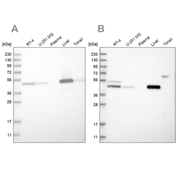
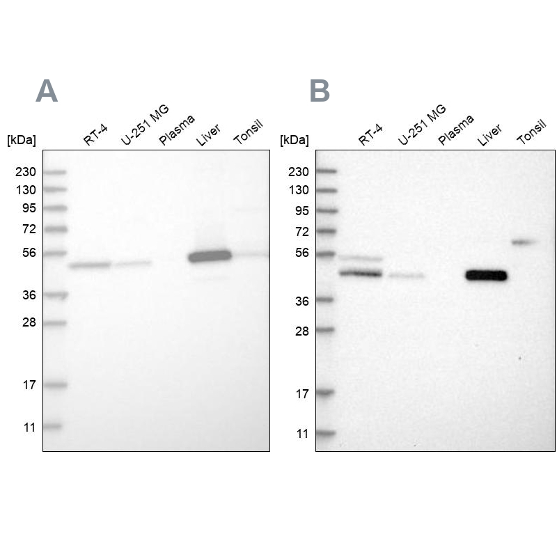
- Experimental details
- Western blot analysis using Anti-PCYT2 antibody HPA023034 (A) shows similar pattern to independent antibody HPA023033 (B).
Supportive validation
- Submitted by
- Atlas Antibodies (provider)
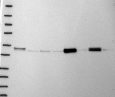
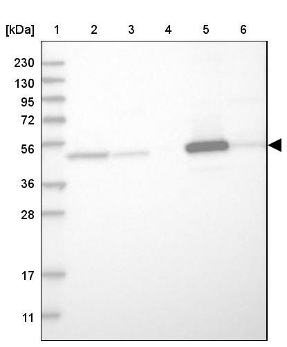
- Main image

- Experimental details
- Lane 1: Marker [kDa] 230, 130, 95, 72, 56, 36, 28, 17, 11Lane 2: Human cell line RT-4Lane 3: Human cell line U-251MG spLane 4: Human plasma (IgG/HSA depleted)Lane 5: Human liver tissueLane 6: Human tonsil tissue
Supportive validation
- Submitted by
- Atlas Antibodies (provider)
- Main image
- Experimental details
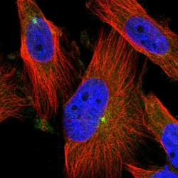
- Immunofluorescent staining of human cell line U-251 MG shows localization to centrosome.
- Sample type
- HUMAN

Enhanced validation
Supportive validation
- Submitted by
- Atlas Antibodies (provider)
- Enhanced method
- Independent antibody validation
- Main image
- Experimental details
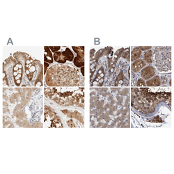
- Immunohistochemical staining of human colon, kidney, liver and testis using Anti-PCYT2 antibody HPA023034 (A) shows similar protein distribution across tissues to independent antibody HPA023033 (B).

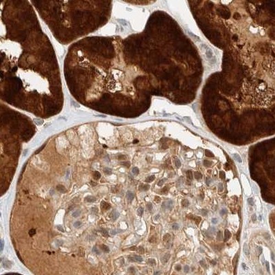
Supportive validation
- Submitted by
- Atlas Antibodies (provider)
- Main image
- Experimental details
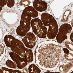
- Immunohistochemical staining of human kidney shows strong cytoplasmic and nuclear positivity in tubular cells.
- Submitted by
- Atlas Antibodies (provider)
- Main image

- Experimental details
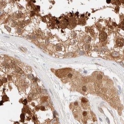
- Immunohistochemical staining of human testis using Anti-PCYT2 antibody HPA023034.
- Sample type
- HUMAN
- Submitted by
- Atlas Antibodies (provider)
- Main image
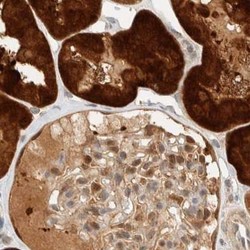
- Experimental details
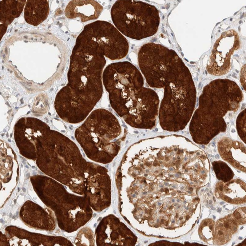
- Immunohistochemical staining of human kidney using Anti-PCYT2 antibody HPA023034.
- Sample type
- HUMAN
- Submitted by
- Atlas Antibodies (provider)
- Main image
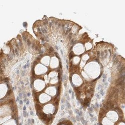
- Experimental details
- Immunohistochemical staining of human colon using Anti-PCYT2 antibody HPA023034.
- Sample type
- HUMAN

- Submitted by
- Atlas Antibodies (provider)
- Main image
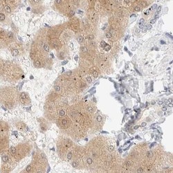
- Experimental details
- Immunohistochemical staining of human liver using Anti-PCYT2 antibody HPA023034.
- Sample type
- HUMAN
