 Explore
 Validate
 Learn
Learn14410-1-AP
antibody from Invitrogen Antibodies
Targeting: TAB2
KIAA0733, MAP3K7IP2
 Western blot Immunocytochemistry
 Immunoprecipitation Immunohistochemistry Flow cytometry Other assay
Antibody data
- Antibody Data
- Antigen structure
- References [0]
- Comments [0]
- Validations
- Western blot [4]
- Immunocytochemistry [1]
- Immunohistochemistry [2]
- Flow cytometry [1]
- Other assay [1]
Submit
Validation data
Reference
Comment
Report error
- Product number
- 14410-1-AP - Provider product page

- Provider
- Invitrogen Antibodies
- Product name
- TAB2 Polyclonal Antibody
- Antibody type
- Polyclonal
- Antigen
- Other
- Description
- Immunogen sequence: SGPRTSSTS SSVNSQTLNR NQPTVYIAAS PPNTDELMSR SQPKVYISAN AATGDEQVMR NQPTLFISTN SGASAASRNM SGQVSMGPAF IHHHPPKSRA IGNNSATSPR VVVTQPNTKY TFKITVSPNK PPAVSPGVVS PTFELTNLLN HPDHYVETEN IQHLTDPTLA HVDRISETRK LSMGSDDAAY TQALLVHQKA RMERLQRELE IQKKKLDKLK SEVNEMENNL TRRRLKRSNS ISQIPSLEEM QQLRSCNRQL QIDIDCLTKE IDLFQARGPH FNPSAIHNFY DNIGFVGPVP PKPKDQRSII KTPKTQDTED DEGAQWNCTA CTFLNHPALI RCEQCEMPRH F (344-693 aa encoded by BC035910)
- Reactivity
- Human, Mouse, Rat
- Host
- Rabbit
- Isotype
- IgG
- Vial size
- 150 µL
- Concentration
- 0.21 mg/mL
- Storage
- -20°C
No comments: Submit comment
Supportive validation
- Submitted by
- Invitrogen Antibodies (provider)
- Main image

- Experimental details
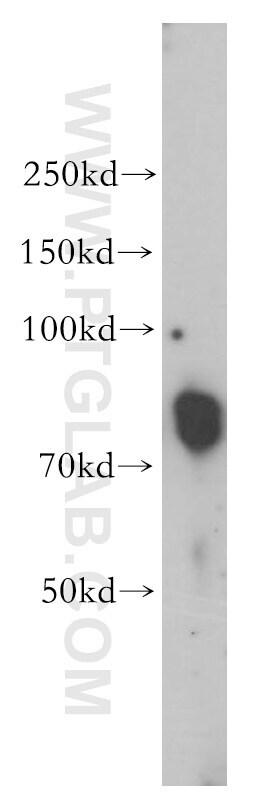
- Human liver tissue were subjected to SDS PAGE followed by western blot with 14410-1-AP (TAB2 antibody) at dilution of 1:300 incubated at room temperature for 1.5 hours.
- Submitted by
- Invitrogen Antibodies (provider)
- Main image

- Experimental details
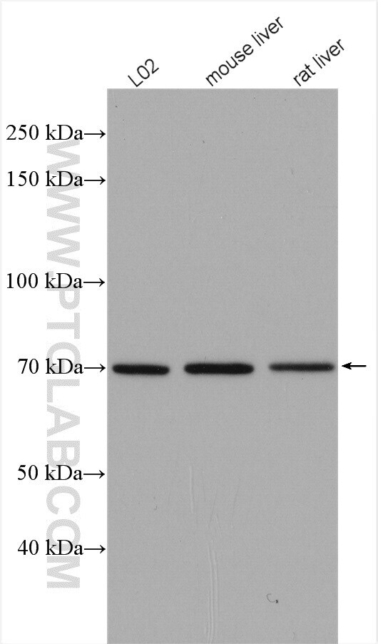
- Various lysates were subjected to SDS PAGE followed by western blot with 14410-1-AP (TAB2 antibody) at dilution of 1:2000 incubated at room temperature for 1.5 hours.
- Submitted by
- Invitrogen Antibodies (provider)
- Main image

- Experimental details
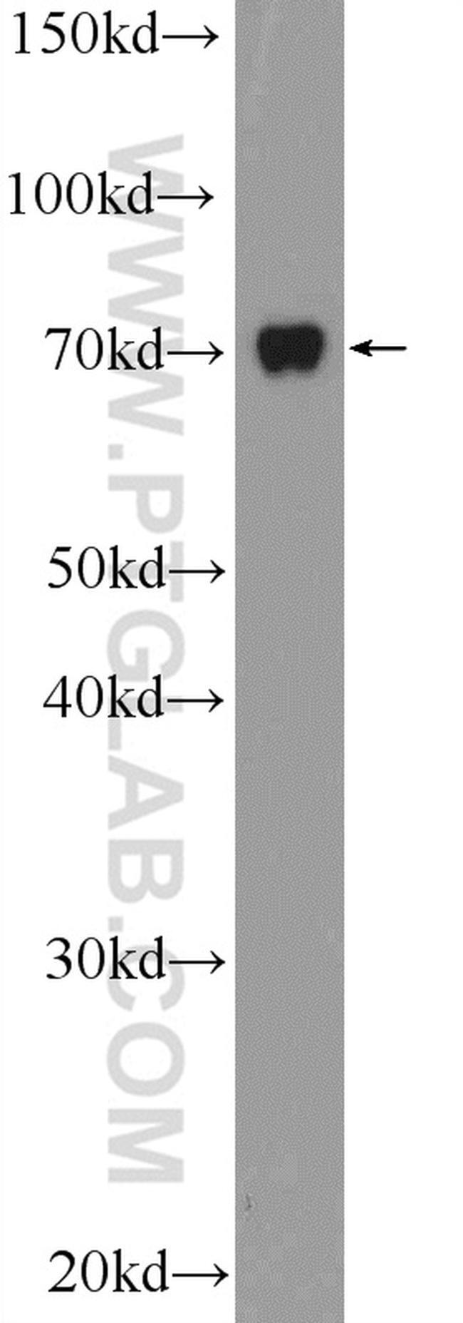
- HepG2 cells were subjected to SDS PAGE followed by western blot with 14410-1-AP ( TAB2 antibody at dilution of 1:600 incubated at room temperature for 1.5 hours.
- Submitted by
- Invitrogen Antibodies (provider)
- Main image

- Experimental details
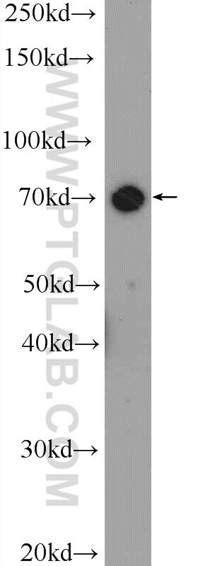
- L02 cells were subjected to SDS PAGE followed by western blot with 14410-1-AP (TAB2 antibody at dilution of 1:600 incubated at room temperature for 1.5 hours.
Supportive validation
- Submitted by
- Invitrogen Antibodies (provider)
- Main image
- Experimental details
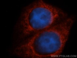
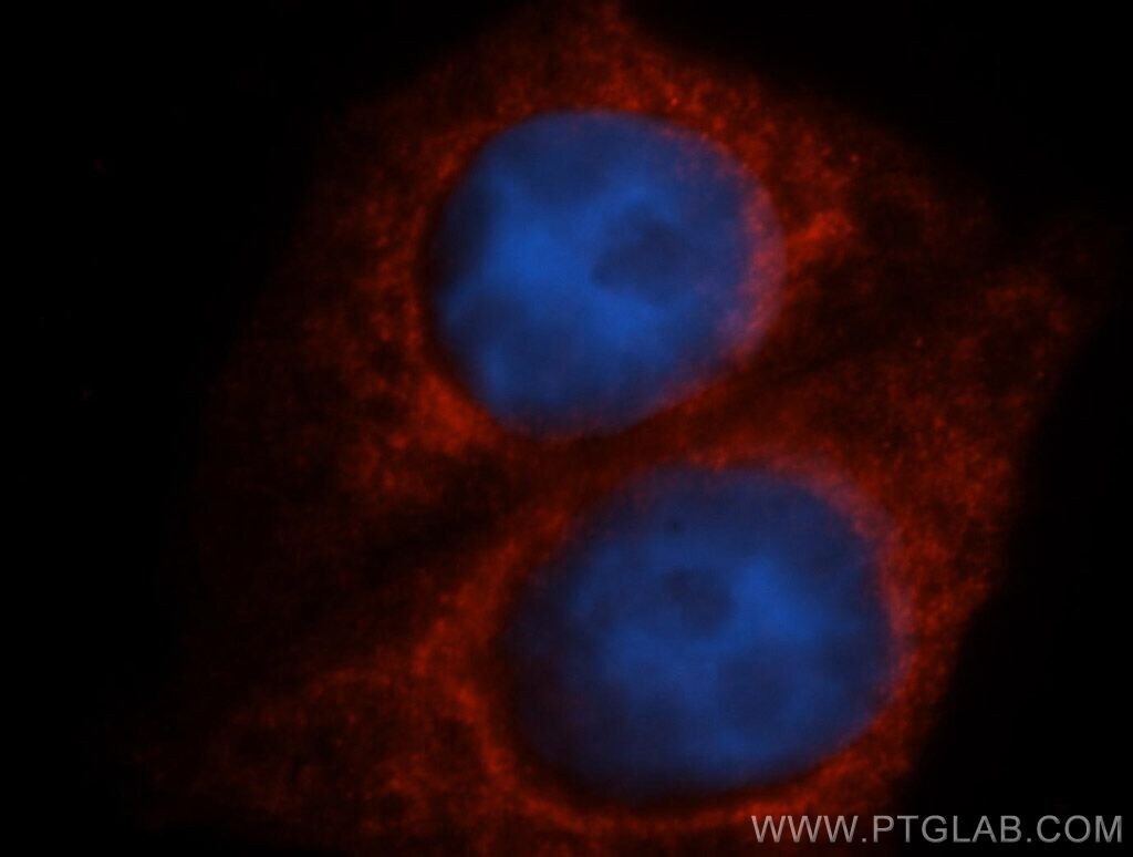
- Immunofluorescent analysis of HepG2 cells, using MAP3K7IP2 antibody 14410-1-AP at 1:50 dilution and Rhodamine-labeled goat anti-rabbit IGG (red). Blue pseudocolor = DAPI (fluorescent DNA dye).
Supportive validation
- Submitted by
- Invitrogen Antibodies (provider)
- Main image

- Experimental details
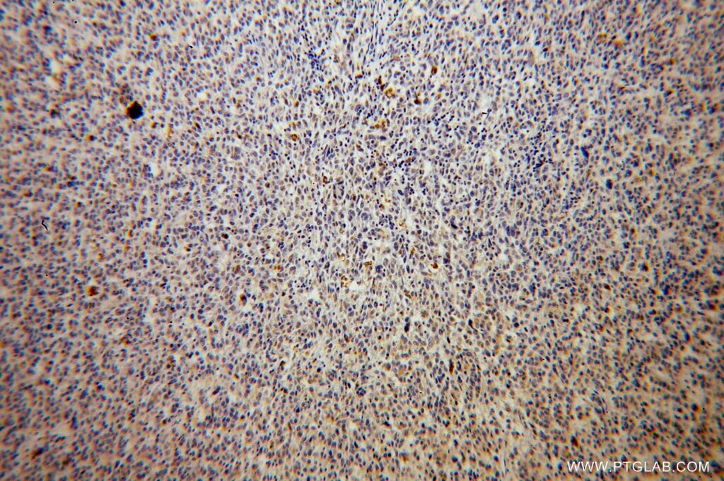
- Immunohistochemistry of paraffin-embedded human malignant melanoma using 14410-1-AP (TAB2 antibody) at dilution of 1:100 (under 10x lens).
- Submitted by
- Invitrogen Antibodies (provider)
- Main image

- Experimental details
- Immunohistochemistry of paraffin-embedded human malignant melanoma using 14410-1-AP (TAB2 antibody) at dilution of 1:100 (under 40x lens).

Supportive validation
- Submitted by
- Invitrogen Antibodies (provider)
- Main image

- Experimental details
- 1X10^6 HepG2 cells were stained with 0.2ug TAB2 antibody (14410-1-AP, red) and control antibody (blue). Fixed with 90% MeOH blocked with 3% BSA (30 min). Alexa Fluor 488-conjugated AffiniPure Goat Anti-Rabbit IGG (H+L) with dilution 1:1000.

Supportive validation
- Submitted by
- Invitrogen Antibodies (provider)
- Main image
- Experimental details
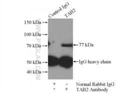
- IP result of anti-TAB2 (IP:14410-1-AP, 4ug; Detection:14410-1-AP 1:600) with HepG2 cells lysate 3600ug.
