Antibody data
- Antibody Data
- Antigen structure
- References [1]
- Comments [0]
- Validations
- Immunohistochemistry [3]
- Other assay [2]
Submit
Validation data
Reference
Comment
Report error
- Product number
- PA5-50864 - Provider product page

- Provider
- Invitrogen Antibodies
- Product name
- WDR45B Polyclonal Antibody
- Antibody type
- Polyclonal
- Antigen
- Synthetic peptide
- Description
- The antibody detects endogenous levels of total WDR45B protein.
- Reactivity
- Human
- Host
- Rabbit
- Isotype
- IgG
- Vial size
- 100 µL
- Concentration
- 2.5 mg/mL
- Storage
- -20°C
Submitted references Wipi3 is essential for alternative autophagy and its loss causes neurodegeneration.
Yamaguchi H, Honda S, Torii S, Shimizu K, Katoh K, Miyake K, Miyake N, Fujikake N, Sakurai HT, Arakawa S, Shimizu S
Nature communications 2020 Oct 20;11(1):5311
Nature communications 2020 Oct 20;11(1):5311
No comments: Submit comment
Supportive validation
- Submitted by
- Invitrogen Antibodies (provider)
- Main image

- Experimental details
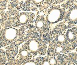
- Immunohistochemical analysis of WDR45B in paraffin-embedded Human thyroid cancer tissue. Samples were probed using a WDR45B polyclonal antibody (Product # PA5-50864) at a dilution of 1/40.
- Submitted by
- Invitrogen Antibodies (provider)
- Main image

- Experimental details
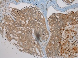
- Immunohistochemical analysis of WDR45B in paraffin embedded Human cervical cancer tissue using WDR45B Polyclonal Antibody (Product # PA5-50864) at a 1:200 dilution.
- Submitted by
- Invitrogen Antibodies (provider)
- Main image

- Experimental details
- Immunohistochemical analysis of WDR45B in paraffin embedded Human Liver cancer tissue using WDR45B Polyclonal Antibody (Product # PA5-50864) at a 1:200 dilution.
Supportive validation
- Submitted by
- Invitrogen Antibodies (provider)
- Main image

- Experimental details
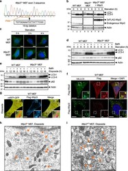
- Fig. 6 Comparison of the contribution of Wipi3 and Wipi2 to canonical autophagy. a Wipi3 Cr MEFs were generated from WT MEFs using the CRISPR/Cas9 system. A 19-bp deletion of wipi3 (third exon) was confirmed by genomic sequencing. The deleted nucleotide sequence and the protospacer adjacent motif (PAM) are indicated below the sequence. b The indicated MEFs were starved for the indicated times, and the expression of the indicated proteins was analyzed by western blotting. Actin was included as a loading control. c The indicated MEFs expressing GFP-LC3 were starved for the indicated times, and GFP-LC3 puncta formation was analyzed. Representative images are shown. Bars = 10 um. d The indicated MEFs were starved with or without bafilomycin A1 (10 nM) for the indicated times, and the expression of each protein was analyzed by western blotting. e Similar experiments to ( d ) were performed using etoposide (10 µM), instead of starvation. The deletion of wipi3 showed a minimal effect on canonical autophagy. f Flag-Wipi2 or Flag-Wipi3 were expressed in HA-LC3-expressing WT MEFs, and were treated with etoposide (10 µM) for 12 h. Then, cells were immunostained with anti-HA and anti-Flag antibodies. Bars = 10 um. Magnified images of the dashed squares are shown in the insets. Bars = 2 um. LC3 signals were merged with Wipi2 but not with Wipi3. g Flag-Wipi3 and mRFP-GFP were expressed in WT MEFs, and were treated with etoposide (10 µM) for 12 h. Then, cells were immunost
- Submitted by
- Invitrogen Antibodies (provider)
- Main image

- Experimental details
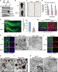
- Fig. 7 Neurological defects in neuron-specific Wipi3 cKO mice. a Western blot analysis of brain lysates. The asterisk indicates a nonspecific band. Wipi3 cKO mice at 18-weeks were compared with Atg7 cKO mice at 5-weeks, as they showed equivalent disease severity. b - d Abnormal motor performance in Wipi3 cKO mice at 10-weeks. The limb-clasping reflex was observed ( b ). The footprint assay indicated a motor deficit ( c ). In ( d ), the time the indicated mice remained on the rod was measured. Data are shown as the mean +- SD ( n = 3). e - g Cryosections of the indicated cerebellum were immunostained for the Purkinje cell marker calbindin (green), the glial cell marker GFAP (red), and DAPI (blue). Bars = 200 um ( e ) and 20 um ( g ). In ( f ), the number of Purkinje cells in the first fissure of the cerebellum is shown. Data are shown as the mean +- SD ( n = 3). h - m Comparisons of morphology between WT, Wipi3 cKO mice (10-weeks), and Atg7 cKO mice (5-weeks). In ( h ), cryosections of the cerebellum immunostained with anti-calbindin and anti-p62 antibodies. Bar = 10 um. ( i ) Morphology of Golgi in cerebella. Small fragmented and swollen rod-shaped Golgi membranes were observed in the Purkinje cells from Wipi3 cKO mice. Bars = 0.5 um. j Immunostaining of calbindin and ubiquitin (Ub) using cryosections of the cerebellum. Bars = 20 um. k , l The accumulation of fibrils was observed in Purkinje cells from Wipi3 cKO mice. Bars = 0.5 um. Orange and red arrows indicate naked fibril
 Explore
 Validate
 Learn
Learn Immunohistochemistry