 Explore
 Validate
 Learn
LearnOAAB05713
antibody from Aviva Systems Biology
Targeting: CFLAR
c-FLIP, CASH, CASP8AP1, Casper, cFLIP, CLARP, FLAME, FLIP, I-FLICE, MRIT
 Western blot
 ELISA
ELISAAntibody data
- Antibody Data
- Antigen structure
- References [3]
- Comments [0]
- Validations
- Western blot [1]
- Immunohistochemistry [1]
- Flow cytometry [1]
Submit
Validation data
Reference
Comment
Report error
- Product number
- OAAB05713 - Provider product page

- Provider
- Aviva Systems Biology
- Product name
- CFLAR antibody - center region
- Antibody type
- Polyclonal
- Reactivity
- Human
- Host
- Rabbit
- Vial size
- 400ul
- Storage
- Maintain refrigerated at 2-8 deg C for up to 6 months. For long term storage store at -20 deg C in small aliquots to prevent freeze-thaw cycles.
Submitted references Release of an ~55kDa fragment containing the actin-binding domain of β-spectrin by caspase-8 during FND-induced apoptosis depends on the presence of protein 4.1.
Activation by C5a of endothelial cell caspase 8 and cFLIP.
Resistance to cytotoxic chemotherapy-induced apoptosis in side population cells of human oral squamous cell carcinoma cell line Ho-1-N-1.
Toporkiewicz M, Grzybek M, Meissner J, Michalczyk I, Dubielecka PM, Korycka J, Seweryn E, Sikorski AF
Archives of biochemistry and biophysics 2013 Jul 15;535(2):205-13
Archives of biochemistry and biophysics 2013 Jul 15;535(2):205-13
Activation by C5a of endothelial cell caspase 8 and cFLIP.
Albrecht EA, Sarma JV, Ward PA
Inflammation research : official journal of the European Histamine Research Society ... [et al.] 2009 Jan;58(1):30-7
Inflammation research : official journal of the European Histamine Research Society ... [et al.] 2009 Jan;58(1):30-7
Resistance to cytotoxic chemotherapy-induced apoptosis in side population cells of human oral squamous cell carcinoma cell line Ho-1-N-1.
Yajima T, Ochiai H, Uchiyama T, Takano N, Shibahara T, Azuma T
International journal of oncology 2009 Aug;35(2):273-80
International journal of oncology 2009 Aug;35(2):273-80
No comments: Submit comment
Supportive validation
- Submitted by
- Aviva Systems Biology (provider)
- Main image

- Experimental details
- Western blot analysis of CFLAR Antibody (Center) in HepG2 cell lysates (35ug/lane). CFLAR (arrow) was detected using the purified Pab.
- Sample type
- HepG2 cell line lysates
- Primary Ab dilution
- 1.0 µg/mL
- Protocol
- Protocol

Supportive validation
- Submitted by
- Aviva Systems Biology (provider)
- Main image

- Experimental details
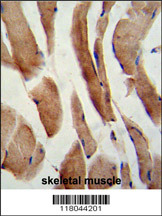
- Formalin-fixed and paraffin-embedded human skeletal muscle reacted with CFLAR Antibody (Center), which was peroxidase-conjugated to the secondary antibody, followed by DAB staining. This data demonstrates the use of this antibody for immunohistochemistry; clinical relevance has not been evaluated.
- Sample type
- human skeletal muscle
- Primary Ab dilution
- 1.0 µg/mL
- Protocol
- Protocol
Supportive validation
- Submitted by
- Aviva Systems Biology (provider)
- Main image

- Experimental details
- CFLAR Antibody (Center) flow cytometric analysis of k562 cells (bottom histogram) compared to a negative control cell (top histogram).FITC-conjugated goat-anti-rabbit secondary antibodies were used for the analysis.
- Sample type
- k562 cells
- Primary Ab dilution
- 1.0 µg/mL
- Protocol
- Protocol
