Explore
 Validate
 Learn
Learn Western blot
Antibody data
- Antibody Data
- Antigen structure
- References [2]
- Comments [0]
- Validations
- Western blot [1]
- Immunocytochemistry [1]
- Flow cytometry [1]
Submit
Validation data
Reference
Comment
Report error
- Product number
- AF-386-PB - Provider product page

- Provider
- R&D Systems
- Product name
- Human CTLA-4 Antibody
- Antibody type
- Polyclonal
- Description
- Antigen Affinity-purified. Detects human CTLA-4 in direct ELISAs and Western blots. In Western blots, approximately 25% cross-reactivity with recombinant mouse CTLA-4 is observed.
- Reactivity
- Human
- Host
- Goat
- Conjugate
- Unconjugated
- Antigen sequence
Q6GR94- Isotype
- IgG
- Vial size
- 100 ug
- Concentration
- LYOPH
- Storage
- Use a manual defrost freezer and avoid repeated freeze-thaw cycles. 12 months from date of receipt, -20 to -70 °C as supplied. 1 month, 2 to 8 °C under sterile conditions after reconstitution. 6 months, -20 to -70 °C under sterile conditions after reconstitution.
Submitted references PD-L1 expression and its relationship with oncogenic drivers in non-small cell lung cancer (NSCLC).
Inhibitory receptors are expressed by Trypanosoma cruzi-specific effector T cells and in hearts of subjects with chronic Chagas disease.
Jiang L, Su X, Zhang T, Yin X, Zhang M, Fu H, Han H, Sun Y, Dong L, Qian J, Xu Y, Fu X, Gavine PR, Zhou Y, Tian K, Huang J, Shen D, Jiang H, Yao Y, Han B, Gu Y
Oncotarget 2017 Apr 18;8(16):26845-26857
Oncotarget 2017 Apr 18;8(16):26845-26857
Inhibitory receptors are expressed by Trypanosoma cruzi-specific effector T cells and in hearts of subjects with chronic Chagas disease.
Argüello RJ, Albareda MC, Alvarez MG, Bertocchi G, Armenti AH, Vigliano C, Meckert PC, Tarleton RL, Laucella SA
PloS one 2012;7(5):e35966
PloS one 2012;7(5):e35966
No comments: Submit comment
Supportive validation
- Submitted by
- R&D Systems (provider)
- Main image
- Experimental details
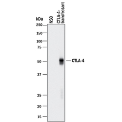
- Detection of Human CTLA-4 by Western Blot. Western blot shows lysates of NS0 mouse myeloma cell line either mock transfected or transfected with human CTLA-4. PVDF membrane was probed with 0.5 µg/mL of Goat Anti-Human CTLA-4 Antigen Affinity-purified Polyclonal Antibody (Catalog # AF-386-PB) followed by HRP-conjugated Anti-Goat IgG Secondary Antibody (Catalog # HAF017). A specific band was detected for CTLA-4 at approximately 50 kDa (as indicated). This experiment was conducted under reducing conditions and using Immunoblot Buffer Group 1.

Supportive validation
- Submitted by
- R&D Systems (provider)
- Main image

- Experimental details
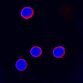
- CTLA-4 in Human Peripheral Blood Mononuclear Cells. CTLA-4 was detected in immersion fixed human peripheral blood mononuclear cells treated with treated with PMA and calcium ionomycin using Goat Anti-Human CTLA-4 Antigen Affinity-purified Polyclonal Antibody (Catalog # AF-386-PB) at 15 µg/mL for 3 hours at room temperature. Cells were stained using the NorthernLights™ 557-conjugated Anti-Goat IgG Secondary Antibody (red; Catalog # NL001) and counterstained with DAPI (blue). Specific staining was localized to cell surfaces. View our protocol for Fluorescent ICC Staining of Non-adherent Cells.
Supportive validation
- Submitted by
- R&D Systems (provider)
- Main image

- Experimental details
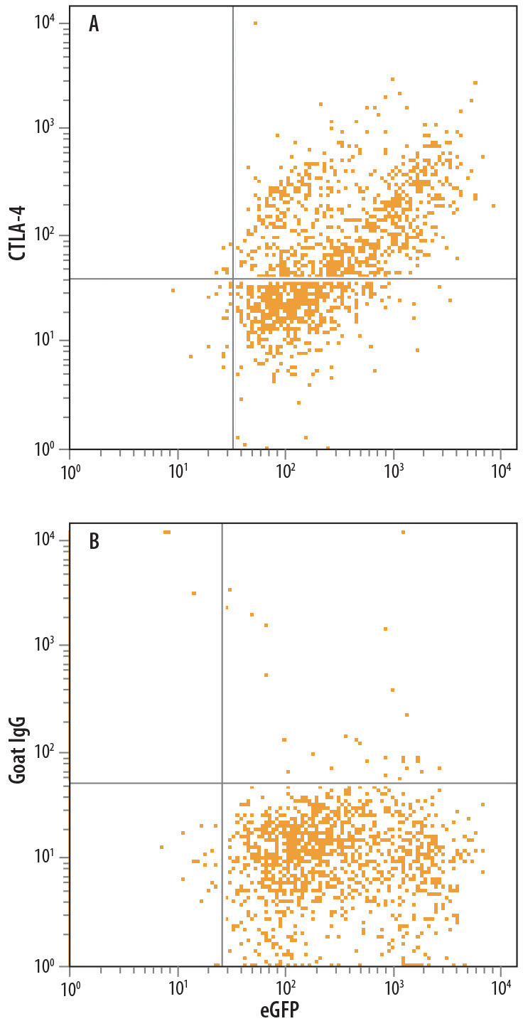
- Detection of CTLA-4 in NS0 Mouse Cell Line Co-transfected with CTLA-4 and eGFP by Flow Cytometry. NS0 mouse myeloma cell line co-transfected with human CTLA-4 and eGFP was stained with either (A) Goat Anti-Human CTLA-4 Antigen Affinity-purified Polyclonal Antibody (Catalog # AF-386-PB) or (B) Normal Goat IgG Control (Catalog # AB-108-C) followed by Allophycocyanin-conjugated Anti-Goat IgG Secondary Antibody (Catalog # F0108).