Explore
 Validate
 Learn
Learn Western blot
Antibody data
- Antibody Data
- Antigen structure
- References [1]
- Comments [0]
- Validations
- Western blot [1]
- Immunocytochemistry [1]
- Other assay [1]
Submit
Validation data
Reference
Comment
Report error
- Product number
- PA5-17295 - Provider product page

- Provider
- Invitrogen Antibodies
- Product name
- PSMA5 Polyclonal Antibody
- Antibody type
- Polyclonal
- Antigen
- Synthetic peptide
- Description
- It is not recommended to aliquot this antibody.
- Reactivity
- Human, Mouse, Rat
- Host
- Rabbit
- Isotype
- IgG
- Vial size
- 100 µL
- Storage
- -20°C
Submitted references SIVcol Nef counteracts SERINC5 by promoting its proteasomal degradation but does not efficiently enhance HIV-1 replication in human CD4+ T cells and lymphoid tissue.
Kmiec D, Akbil B, Ananth S, Hotter D, Sparrer KMJ, Stürzel CM, Trautz B, Ayouba A, Peeters M, Yao Z, Stagljar I, Passos V, Zillinger T, Goffinet C, Sauter D, Fackler OT, Kirchhoff F
PLoS pathogens 2018 Aug;14(8):e1007269
PLoS pathogens 2018 Aug;14(8):e1007269
No comments: Submit comment
Supportive validation
- Submitted by
- Invitrogen Antibodies (provider)
- Main image
- Experimental details
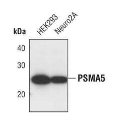
- Western blot analysis of PSMA5 in extracts from HEK293 and Neuro2A cell lines using PSMA5 polyclonal antibody (Product # PA5-17295).

Supportive validation
- Submitted by
- Invitrogen Antibodies (provider)
- Main image
- Experimental details
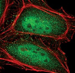
- Immunofluorescent analysis of PSMA5 in HeLa cells using a PSMA5 polyclonal antibody (Product # PA5-17295) (green). Actin filaments are labeled with a fluorescent red phalloidin.

Supportive validation
- Submitted by
- Invitrogen Antibodies (provider)
- Main image
- Experimental details
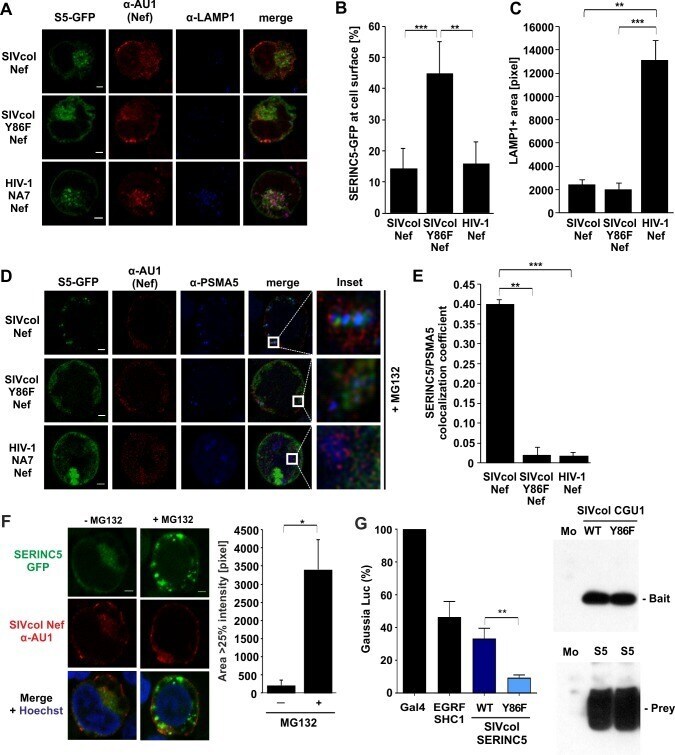
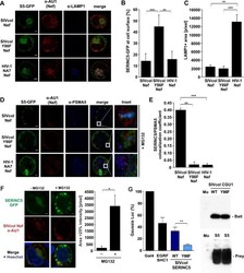
- Fig 7 SIVcol Nef relocalizes SERINC5 to the proteasome by an Y86 dependent mechanism. (A) Representative Laser scanning confocal microscopy images of JTAg SERINC3/5 knock-out cells transfected with SERINC5-GFP (S5-GFP; green) alone or together with the indicated AU1-tagged Nef proteins (red). Endogenous lysosomes were stained with an anti-LAMP1 antibody (blue). (B) Quantification of internal SERINC-GFP fluorescence versus surface SERINC-GFP fluorescence from (A) displayed as means of n = 5 (+SD). (C) Quantification of the pixel area of LAMP1 staining from (A) in triplicates (+SD). (D) Representative Laser scanning confocal microscopy images of JTAg SERINC3/5 knock-out cells transfected with SERINC5-GFP (green) together with indicated AU1-tagged Nef (red) and endogenous proteasomes (anti-PSMA5, blue). Insets show magnifications of the highlighted areas. Size bar, 2 mum. (E) Calculation of Pearson's co-localization coefficients using Costes thresholds for PSMA5 (proteasome) and SERINC5-GFP for images in (D). Displayed as means of triplicates (+SD). (F) Representative Laser scanning confocal microscopy images (left) and quantitative analysis (right) of JTAg SERINC3/5 knock-out cells transfected with SERINC5-GFP (green) together with AU1-tagged SIVcol Nef wt (red) and either treated with MG132 (10 muM for 3 h) or mock treated. Nuclei are stained with Hoechst (blue). (G) HEK293T B0166 MaMTH reporter cells were co-transfected with 25 ng Bait (Nef) and 25 ng Prey (SERINC5) DNA. Aft