Antibody data
- Antibody Data
- Antigen structure
- References [4]
- Comments [0]
- Validations
- Western blot [1]
- Immunocytochemistry [3]
- Immunohistochemistry [3]
- Other assay [2]
Submit
Validation data
Reference
Comment
Report error
- Product number
- MA5-32067 - Provider product page

- Provider
- Invitrogen Antibodies
- Product name
- NGF Recombinant Rabbit Monoclonal Antibody (SI79-01)
- Antibody type
- Monoclonal
- Antigen
- Synthetic peptide
- Description
- Recombinant rabbit monoclonal antibodies are produced using in vitro expression systems. The expression systems are developed by cloning in the specific antibody DNA sequences from immunoreactive rabbits. Then, individual clones are screened to select the best candidates for production. The advantages of using recombinant rabbit monoclonal antibodies include: better specificity and sensitivity, lot-to-lot consistency, animal origin-free formulations, and broader immunoreactivity to diverse targets due to larger rabbit immune repertoire.
- Reactivity
- Human, Mouse, Rat, Zebrafish
- Host
- Rabbit
- Isotype
- IgG
- Antibody clone number
- SI79-01
- Vial size
- 100 µL
- Concentration
- 1 mg/mL
- Storage
- Store at 4°C short term. For long term storage, store at -20°C, avoiding freeze/thaw cycles.
Submitted references 4-Aminopyridine Induces Nerve Growth Factor to Improve Skin Wound Healing and Tissue Regeneration.
Resveratrol Inhibition of the WNT/β-Catenin Pathway following Discogenic Low Back Pain.
Synergic Neuroprotection Between Ligusticum Chuanxiong Hort and Borneol Against Ischemic Stroke by Neurogenesis via Modulating Reactive Astrogliosis and Maintaining the Blood-Brain Barrier.
Circulating microRNAs Profile in Patients With Transthyretin Variant Amyloidosis.
Jagadeeshaprasad MG, Govindappa PK, Nelson AM, Noble MD, Elfar JC
Biomedicines 2022 Jul 8;10(7)
Biomedicines 2022 Jul 8;10(7)
Resveratrol Inhibition of the WNT/β-Catenin Pathway following Discogenic Low Back Pain.
Genovese T, Impellizzeri D, D'Amico R, Cordaro M, Peritore AF, Crupi R, Gugliandolo E, Cuzzocrea S, Fusco R, Siracusa R, Di Paola R
International journal of molecular sciences 2022 Apr 7;23(8)
International journal of molecular sciences 2022 Apr 7;23(8)
Synergic Neuroprotection Between Ligusticum Chuanxiong Hort and Borneol Against Ischemic Stroke by Neurogenesis via Modulating Reactive Astrogliosis and Maintaining the Blood-Brain Barrier.
Yu B, Yao Y, Zhang X, Ruan M, Zhang Z, Xu L, Liang T, Lu J
Frontiers in pharmacology 2021;12:666790
Frontiers in pharmacology 2021;12:666790
Circulating microRNAs Profile in Patients With Transthyretin Variant Amyloidosis.
Vita GL, Aguennouz M, Polito F, Oteri R, Russo M, Gentile L, Barbagallo C, Ragusa M, Rodolico C, Di Giorgio RM, Toscano A, Vita G, Mazzeo A
Frontiers in molecular neuroscience 2020;13:102
Frontiers in molecular neuroscience 2020;13:102
No comments: Submit comment
Supportive validation
- Submitted by
- Invitrogen Antibodies (provider)
- Main image

- Experimental details
- Western blot analysis of NGF in Hela cell lysate using a NGF Monoclonal antibody (Product # MA5-32067) at a dilution of 1:1,000.
Supportive validation
- Submitted by
- Invitrogen Antibodies (provider)
- Main image

- Experimental details
- Immunocytochemical analysis of NGF in HepG2 cells using a NGF Monoclonal antibody (Product # MA5-32067) as seen in green. The nuclear counter stain is DAPI (blue). Cells were fixed in paraformaldehyde, permeabilised with 0.25% Triton X100/PBS.
- Submitted by
- Invitrogen Antibodies (provider)
- Main image

- Experimental details
- Immunocytochemical analysis of NGF in NIH/3T3 cells using a NGF Monoclonal antibody (Product # MA5-32067) as seen in green. The nuclear counter stain is DAPI (blue). Cells were fixed in paraformaldehyde, permeabilised with 0.25% Triton X100/PBS.
- Submitted by
- Invitrogen Antibodies (provider)
- Main image

- Experimental details
- Immunocytochemical analysis of NGF in Hela cells using a NGF Monoclonal antibody (Product # MA5-32067) as seen in green. The nuclear counter stain is DAPI (blue). Cells were fixed in paraformaldehyde, permeabilised with 0.25% Triton X100/PBS.
Supportive validation
- Submitted by
- Invitrogen Antibodies (provider)
- Main image

- Experimental details
- Immunohistochemical analysis of NGF of paraffin-embedded Mouse liver tissue using a NGF Monoclonal antibody (Product #MA5-32067). Counter stained with hematoxylin.
- Submitted by
- Invitrogen Antibodies (provider)
- Main image

- Experimental details
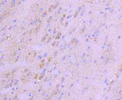
- Immunohistochemical analysis of NGF of paraffin-embedded Mouse brain tissue using a NGF Monoclonal antibody (Product #MA5-32067). Counter stained with hematoxylin.
- Submitted by
- Invitrogen Antibodies (provider)
- Main image

- Experimental details
- Immunohistochemical analysis of NGF of paraffin-embedded Mouse thymus tissue using a NGF Monoclonal antibody (Product #MA5-32067). Counter stained with hematoxylin.
Supportive validation
- Submitted by
- Invitrogen Antibodies (provider)
- Main image

- Experimental details
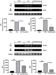
- Resveratrol administration reduced pain-related signaling. Western blot analysis of: trkA ( A ), NGF ( B ) expression in the disc tissue; Western blot analysis of: trkA ( C ), NGF ( D ) expression in the spinal cord tissue. * p < 0.05 vs. CFA, ## p < 0.01 vs. sham, ** p < 0.01 vs. CFA, ### p < 0.001 vs. sham, *** p < 0.001 vs. CFA. Error bar shown as SEM.
- Submitted by
- Invitrogen Antibodies (provider)
- Main image

- Experimental details
- Figure 6 4-AP enhanced expression of transcription factors, neurotrophic factors, and neuropeptides associated with reinnervation. ( a ) Representative image of triple co-immunostaining of wound skin for the transcription factor SOX10 (green), neuropeptide substance-P (SP--yellow), nerve growth factor (NGF--red), and nuclear stain DAPI (blue) and dashed line denotes epidermal/dermal border. Scale bars = 50 mum. ( b - d ) Quantification of SOX10, substance-P, and NGF expressing cells in healed wounds. Data represents 20 images from 5 different mouse wounds and are shown as mean +- SEM, n = 5 animals per group, statistical significance indicated by asterisks (O = saline mouse data points, = 4-AP treated mouse data points, * = p between 0.01 and 0.05, ** = p between 0.01 and 0.001, and *** = p between 0.001 and 0.0002 vs. saline). ( e ) Representative image of Western blot of SOX10, NGF, and GAPDH. ( f , g ) Normalized integrated densities for SOX10 and NGF immunoblot. Mean +- SEM, n = 3 animals per group, statistical significance indicated by asterisks (O = saline mouse data points, = 4-AP treated mouse data points, ** = p between 0.01 and 0.001, and *** = p between 0.001 and 0.0002 vs. saline).
 Explore
 Validate
 Learn
Learn Western blot