Explore
 Validate
 Learn
Learn Immunocytochemistry
Antibody data
- Antibody Data
- Antigen structure
- References [3]
- Comments [0]
- Validations
- Immunocytochemistry [1]
- Immunohistochemistry [5]
Submit
Validation data
Reference
Comment
Report error
- Product number
- HPA008436 - Provider product page

- Provider
- Atlas Antibodies
- Proper citation
- Atlas Antibodies Cat#HPA008436, RRID:AB_1850757
- Product name
- Anti-HJURP
- Antibody type
- Polyclonal
- Reactivity
- Human
- Host
- Rabbit
- Conjugate
- Unconjugated
- Antigen sequence
TPVVQMATLTYETPQGLRIWGGRLIKERNEGEIQD
SSMKPADRTDGSVQAAAWGPELPSHRTVLGADSKS
GEVDATSDQEESVAWALAPAVPQSPLKNELRRKYL
TQVDILLQGAEYFECAGNRAGRDVRV- Isotype
- IgG
- Vial size
- 100 µl
- Storage
- Store at +4°C for short term storage. Long time storage is recommended at -20°C.
Submitted references Prognostic Significance of EDN/RB, HJURP, p60/CAF-1 and PDLI4, Four New Markers in High-Grade Gliomas
The expression level of HJURP has an independent prognostic impact and predicts the sensitivity to radiotherapy in breast cancer.
HJURP binds CENP-A via a highly conserved N-terminal domain and mediates its deposition at centromeres
de Tayrac M, Saikali S, Aubry M, Bellaud P, Boniface R, Quillien V, Mosser J, Jiang T
PLoS ONE 2013 September;8(9)
PLoS ONE 2013 September;8(9)
The expression level of HJURP has an independent prognostic impact and predicts the sensitivity to radiotherapy in breast cancer.
Hu Z, Huang G, Sadanandam A, Gu S, Lenburg ME, Pai M, Bayani N, Blakely EA, Gray JW, Mao JH
Breast cancer research : BCR 2010;12(2):R18
Breast cancer research : BCR 2010;12(2):R18
HJURP binds CENP-A via a highly conserved N-terminal domain and mediates its deposition at centromeres
Shuaib M, Ouararhni K, Dimitrov S, Hamiche A
Proceedings of the National Academy of Sciences 2010 January;107(4):1349-1354
Proceedings of the National Academy of Sciences 2010 January;107(4):1349-1354
No comments: Submit comment
Supportive validation
- Submitted by
- Atlas Antibodies (provider)
- Main image
- Experimental details
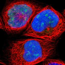
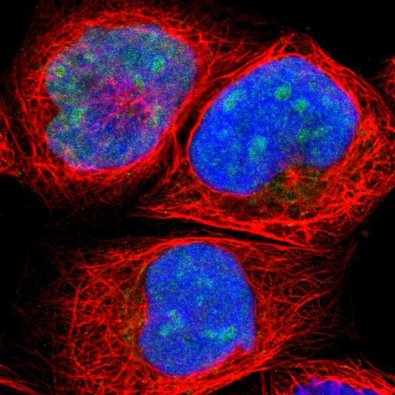
- Immunofluorescent staining of human cell line A-431 shows positivity in nucleus & nucleoli.
- Sample type
- HUMAN
Supportive validation
- Submitted by
- Atlas Antibodies (provider)
- Main image
- Experimental details
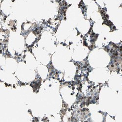
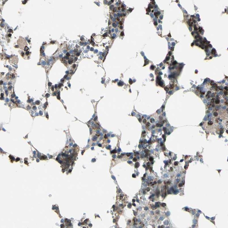
- Immunohistochemical staining of human bone marrow shows strong cytoplasmic positivity in subsets of hematopoietic cells.
- Submitted by
- Atlas Antibodies (provider)
- Main image
- Experimental details
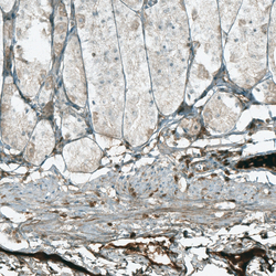
- Immunohistochemical staining of human stomach shows moderate nuclear positivity in lymphoid cells.
- Sample type
- HUMAN
- Submitted by
- Atlas Antibodies (provider)
- Main image

- Experimental details
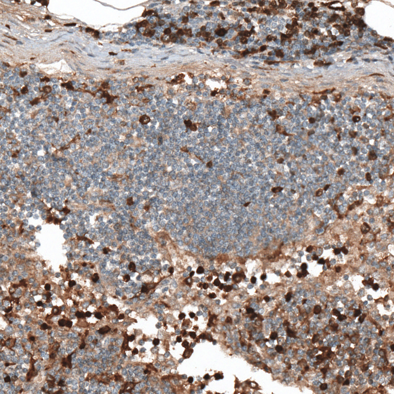
- Immunohistochemical staining of human lymph node shows moderate to strong nuclear positivity in lymphoid cells.
- Sample type
- HUMAN
- Submitted by
- Atlas Antibodies (provider)
- Main image
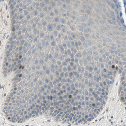
- Experimental details
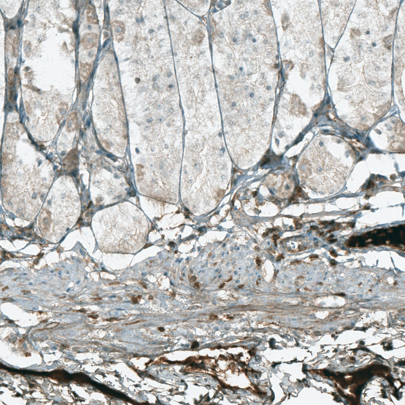
- Immunohistochemical staining of human skin shows moderate nuclear positivity in a subset of basal cells in squamous epithelium.
- Sample type
- HUMAN

- Submitted by
- Atlas Antibodies (provider)
- Main image

- Experimental details
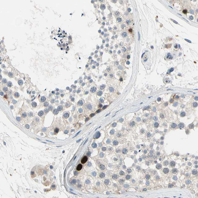
- Immunohistochemical staining of human testis shows moderate to strong nuclear positivity in some spermatogonia cells.
- Sample type
- HUMAN