 Explore
 Validate
 Learn
Learn Western blot
Antibody data
- Antibody Data
- Antigen structure
- References [0]
- Comments [0]
- Validations
- Western blot [2]
- Immunocytochemistry [1]
- Immunohistochemistry [3]
- Flow cytometry [1]
Submit
Validation data
Reference
Comment
Report error
- Product number
- CF506331 - Provider product page

- Provider
- Invitrogen Antibodies
- Product name
- FCGR1A Monoclonal Antibody (OTI1A8), TrueMAB™
- Antibody type
- Monoclonal
- Antigen
- Recombinant full-length protein
- Description
- For reconstitution, we recommend adding 100 µL distilled water to a final antibody concentration of about 1 mg/mL. To use this carrier-free antibody for conjugation experiments, we strongly recommend performing another round of desalting. (Zeba Spin Desalting Columns, 7KMWCO, 0.5 mL, Product # 89882)
- Reactivity
- Human
- Host
- Mouse
- Isotype
- IgG
- Antibody clone number
- OTI1A8
- Vial size
- 100 µg
- Concentration
- 1 mg/mL
- Storage
- -20° C, Avoid Freeze/Thaw Cycles
No comments: Submit comment
Supportive validation
- Submitted by
- Invitrogen Antibodies (provider)
- Main image

- Experimental details
- HEK293T cells were transfected with the pCMV6-ENTRY control (Left lane) or pCMV6-ENTRY FCGR1A (RC207487, Right lane) cDNA for 48 hrs and lysed. Equivalent amounts of cell lysates (5 µg per lane) were separated by SDS-PAGE and immunoblotted with anti-FCGR1. Positive lysates LY400193 (100 µg) and LC400193 (20 µg) can be purchased separately from OriGene.

Supportive validation
- Submitted by
- Invitrogen Antibodies (provider)
- Main image

- Experimental details
- Immunofluorescent staining of 293T cells transfected by pCMV6-ENTRY FCGR1A(RC207487) using anti-FCGR1A antibody (TA506331/green, upper left; DAPI/blue, lower left; MERGED, upper right). 293T cells transfected with empty vector served as a negative control (MERGED, lower right). (1:100)

Supportive validation
- Submitted by
- Invitrogen Antibodies (provider)
- Main image
- Experimental details
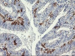
- Immunohistochemical staining of paraffin-embedded Adenocarcinoma of Human endometrium tissue using anti-FCGR1A mouse monoclonal antibody. (Heat-induced epitope retrieval by 10mM citric buffer, pH6.0, 120°C for 3min, TA506331)

- Submitted by
- Invitrogen Antibodies (provider)
- Main image

- Experimental details
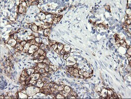
- Immunohistochemical staining of paraffin-embedded Carcinoma of Human prostate tissue using anti-FCGR1A mouse monoclonal antibody. (Heat-induced epitope retrieval by 10mM citric buffer, pH6.0, 120°C for 3min, TA506331)
- Submitted by
- Invitrogen Antibodies (provider)
- Main image

- Experimental details
- Immunohistochemical staining of paraffin-embedded human lymphoma tissue using anti-FCGR1A mouse monoclonal antibody. (Heat-induced epitope retrieval by 10mM citric buffer, pH6.0, 120°C for 3min, TA506331)

Supportive validation
- Submitted by
- Invitrogen Antibodies (provider)
- Main image

- Experimental details
- Flow cytometric analysis of living 293T cells transfected with FCGR1A overexpression plasmid (RC207487), Red)/empty vector (PS100001, Blue) using anti-FCGR1A antibody (TA506331). cells incubated with a non-specific antibody (Green) were used as isotype control. (1:100)
