Antibody data
- Antibody Data
- Antigen structure
- References [0]
- Comments [0]
- Validations
- Western blot [1]
- ELISA [1]
- Immunohistochemistry [1]
- Flow cytometry [1]
Submit
Validation data
Reference
Comment
Report error
- Product number
- DM3511 - Provider product page

- Provider
- Acris Antibodies GmbH
- Proper citation
- Acris Antibodies GmbH Cat#DM3511, RRID:AB_975404
- Product name
- anti CD202b / TEK
- Antibody type
- Monoclonal
- Antigen
- Recombinant Human soluble extracellular domain of TIE-2 protein
- Reactivity
- Human
- Host
- Mouse
- Isotype
- IgG
- Antibody clone number
- Cl.16
- Vial size
- 0.1 mg
No comments: Submit comment
Supportive validation
- Submitted by
- Acris Antibodies GmbH (provider)
- Main image

- Experimental details
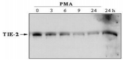
- Efforts of PMA treatment on TIE-2 mRNA and Protein.Lane 1 : HUVECs left untreatedLane 2 : HUVECs stimulated for 3 hours with PMA at 25 ng/mlLane 3 : HUVECs stimulated for 6 hours with PMA at 25 ng/mlLane 4 : HUVECs stimulated for 9 hours with PMA at 25 ng/mlLane 5 : HUVECs stimulated for 24 hours with PMA at 25 ng/ml
Supportive validation
- Submitted by
- Acris Antibodies GmbH (provider)
- Main image

- Experimental details
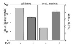
- Quantification of soluble and cellular TIE-2 by Sandwich ELISA: A. CM and cell lysates from HUVECs treated with PMA (25ng/ml) or left untreated were analysed by Sandwich ELISA for the concentrations of sTIE-2 or TIE-2. For Capturing anti-Human TIE-2 Cl.16 (Cat.-No DM3511) was used. for the detection a mixture of biotinylated anti-human TIE-2 Cl.2 (Cat.-No DM3509B) and Cl.9 (Cat.-No DM3510).
Supportive validation
- Submitted by
- Acris Antibodies GmbH (provider)
- Main image

- Experimental details
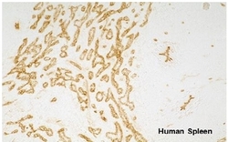
- Immunohistochemistry with Human spleen cryo-sections using monoclonal anti-Human TIE-2 antibody. The experiment was performed by Prof. Dr. Birte Steiniger, Institute of Anatomy and Cell Biology Robert-Koch-Str. 8, D-35037 Marburg, Germany
Supportive validation
- Submitted by
- Acris Antibodies GmbH (provider)
- Main image

- Experimental details
- FACS analysis with human primary lymphatic endothelial cells (HDLEC). As secondary antibody anti-mouse IgG-PE was used.
 Explore
 Validate
 Learn
Learn Western blot