Explore
 Validate
 Learn
LearnNBP1-40256-0.1mg
antibody from Novus Biologicals
Targeting: ATF6
ATF6A
 Western blot
 Immunocytochemistry Immunoprecipitation Immunohistochemistry Flow cytometry Chromatin Immunoprecipitation
Antibody data
- Antibody Data
- Antigen structure
- References [0]
- Comments [0]
- Validations
- Western blot [6]
- Immunohistochemistry [1]
- Flow cytometry [2]
Submit
Validation data
Reference
Comment
Report error
- Product number
- NBP1-40256-0.1mg - Provider product page

- Provider
- Novus Biologicals
- Proper citation
- Novus Cat#NBP1-40256-0.1mg, RRID:AB_11005857
- Product name
- Mouse Monoclonal ATF6 Antibody
- Antibody type
- Monoclonal
- Description
- Protein G purified. This ATF6 antibody detects both the full length and the cleaved/active protein.
- Reactivity
- Human, Mouse, Rat, Porcine, Rabbit
- Host
- Mouse
- Isotype
- IgG
- Vial size
- 0.1 mg
- Concentration
- 1.0 mg/ml
- Storage
- Store at 4C short term. Aliquot and store at -20C long term. Avoid freeze-thaw cycles.
No comments: Submit comment
Supportive validation
- Submitted by
- Novus Biologicals (provider)
- Main image

- Experimental details
- Western Blot: ATF6 Antibody (70B1413.1) [NBP1-40256] - Lane 1: 293 cells transfected with full-length ATF6. Lane 2: 293 cells transfected with partial length ATF6 (amino acids 1-373). Lane 3: Untransfected 293 cells. Western blots were probed with 4 ug/ml of the ATF6 monoclonal antibody and visualized with PicoTect Western Blot Chemiluminescence Substrate (10087K). Film was exposed for 1 min. The top arrow corresponds to the 90 kDa form of ATF6 described as full-length in the literature. The human full-length and partial length ATF6 plasmids are described in Luo and Lee (2002).

- Submitted by
- Novus Biologicals (provider)
- Main image

- Experimental details
- Western Blot: ATF6 Antibody (70B1413.1) [NBP1-40256] - Analysis of ATF6 in mouse liver tissue using 3 ug/ml of ATF6 antibody and 0.25 ug/ml of GAPDH antibody. Lane A contains 20 ugs of whole mouse liver lysate, lane B contains 20 ugs of total ER fraction, and lane C contains 20 ugs of rough ER fraction. The ATF6 band may represent under glycosylated or cleaved/active ATF6.

- Submitted by
- Novus Biologicals (provider)
- Main image
- Experimental details
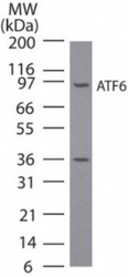
- Western Blot: ATF6 Antibody (70B1413.1) [NBP1-40256] - Analysis of ATF6 in NIH3T3 cell lysate using NBP1-40256 at 3 ug/ml. A band corresponding to full-length ATF6 was detected. We have not characterized the ~36 kDa observed band; it may be an ATF6 breakdown/cleavage product.

- Submitted by
- Novus Biologicals (provider)
- Main image

- Experimental details
- Western Blot: ATF6 Antibody (70B1413.1) [NBP1-40256] - Image of anti-ATF6. Whole cell protein from Hek293 and HepG2 treated with and without 10 mM DTT was separated on a 7.5% gel by SDS-PAGE, transferred to PVDF membrane and blocked in 5% non-fat milk in TBST. The membrane was probed with 5 ug/ml anti-ATF6 and detected with an anti-mouse HRP secondary antibody using a maximum sensitivity ECL reagent. Note the loss of the 90 kDa modified ATF6 protein (arrowhead) upon DTT treatment to activate the unfolded protein response.

- Submitted by
- Novus Biologicals (provider)
- Main image

- Experimental details
- Western Blot: ATF6 Antibody (70B1413.1) [NBP1-40256] - Image of anti-ATF6. Whole cell protein from HeLa cells treated with and without 10 mM DTT was separated on a 7.5% gel by SDS-PAGE, transferred to PVDF membrane and blocked in 5% non-fat milk in TBST. The membrane was probed with 5 ug/ml anti-ATF6 and detected with an anti-mouse HRP secondary antibody using a maximum sensitivity ECL reagent. Note the loss of the 90 kDa modified ATF6 protein (arrowhead) upon DTT treatment to activate the unfolded protein response.

- Submitted by
- Novus Biologicals (provider)
- Main image

- Experimental details
- Western Blot: ATF6 Antibody (70B1413.1) [NBP1-40256] - Hypoxia leads to EMT and ER-stress in CRC cells. B. C. Confluent growing SW480 (B) and HCT116 (C) cells were cultured under conditions of normoxia or hypoxia-like conditions (serum free; 100 uM CoCl2, 1-9 h. B-actin (B-act) as loading control. HIF1a was detectable after 3 h of CoCl2 incubation, the amount of the 50 kD-ATF6 fragment, was already enhanced after 1 h of addition of CoCl2. Image collected and cropped by CiteAb from the following publication (//doi.org/10.1371/journal.pone.0087386) licensed under a CC-BY licence.

Supportive validation
- Submitted by
- Novus Biologicals (provider)
- Main image

- Experimental details
- Immunohistochemistry-Paraffin: ATF6 Antibody (70B1413.1) [NBP1-40256] - Human placenta, followed by biotinylated horse anti-mouse IgG secondary antibody, alkaline phosphatase-streptavidin and chromogen. Dilution 10ug/ml

Supportive validation
- Submitted by
- Novus Biologicals (provider)
- Main image

- Experimental details
- Flow Cytometry: ATF6 Antibody (70B1413.1) [NBP1-40256] - Intracellular flow cytometric staining of 1 x 10^6 MCF-7 cells using ATF6 antibody (dark blue). Isotype control shown in orange. An antibody concentration of 1 ug/1x10^6 cells was used.

- Submitted by
- Novus Biologicals (provider)
- Main image

- Experimental details
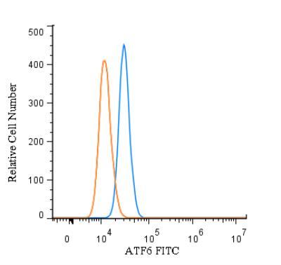
- Flow (Intracellular): ATF6 Antibody (70B1413.1) [NBP1-40256] - An intracellular stain was performed on HeLa cells with ATF6 Antibody (70B1413.1) NBP1-40256F (blue) and a matched isotype control (orange). Cells were fixed with 4% PFA and then permeablized with 0.1% saponin. Cells were incubated in an antibody dilution of 10 ug/mL for 30 minutes at room temperature. Both antibodies were conjugated to FITC.