Explore
 Validate
 Learn
Learn Western blot
Antibody data
- Antibody Data
- Antigen structure
- References [0]
- Comments [0]
- Validations
- Western blot [4]
- Immunohistochemistry [1]
- Other assay [2]
Submit
Validation data
Reference
Comment
Report error
- Product number
- PA5-22332 - Provider product page

- Provider
- Invitrogen Antibodies
- Product name
- RGS4 Polyclonal Antibody
- Antibody type
- Polyclonal
- Antigen
- Recombinant protein fragment
- Description
- Recommended positive controls: Raji, mouse cerebellum, rat brain.
- Concentration
- 0.43 mg/mL
No comments: Submit comment
Supportive validation
- Submitted by
- Invitrogen Antibodies (provider)
- Main image

- Experimental details
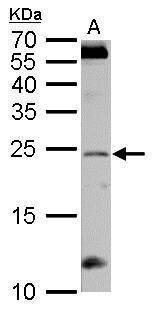
- Western blot analysis of RGS4 using 50 µg mouse cerebellum lysate. Samples were loaded onto a 12% SDS-PAGE gel and probed with a RGS4 polyclonal antibody (Product # PA5-22332) at a dilution of 1:1000.
- Submitted by
- Invitrogen Antibodies (provider)
- Main image
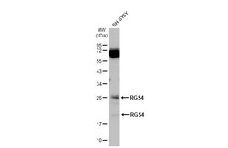
- Experimental details
- Western blot analysis of RGS4 using 30 µg of Raji lysate. Samples were loaded onto a 12% SDS-PAGE gel and probed with a RGS4 polyclonal antibody (Product # PA5-22332) at a dilution of 1:1000.

- Submitted by
- Invitrogen Antibodies (provider)
- Main image
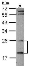
- Experimental details
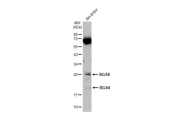
- Western Blot using RGS4 Polyclonal Antibody (Product # PA5-22332). Whole cell extract (30 µg) was separated by 12% SDS-PAGE, and the membrane was blotted with RGS4 Polyclonal Antibody (Product # PA5-22332) diluted at 1:1,000. The HRP-conjugated anti-rabbit IgG antibody was used to detect the primary antibody.
- Submitted by
- Invitrogen Antibodies (provider)
- Main image
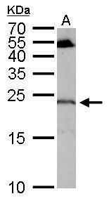
- Experimental details
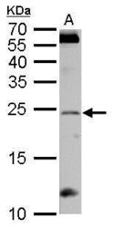
- RGS4 Polyclonal Antibody detects RGS4 protein by western blot analysis. A. 50 µg rat brain lysate/extract.12% SDS-PAGE. RGS4 Polyclonal Antibody (Product # PA5-22332) dilution: 1:1,000. The HRP-conjugated anti-rabbit IgG antibody was used to detect the primary antibody.
Supportive validation
- Submitted by
- Invitrogen Antibodies (provider)
- Main image
- Experimental details
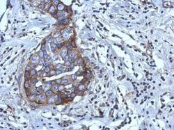
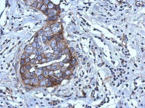
- Immunohistochemical analysis of paraffin-embedded human breast cancer, using RGS4 (Product # PA5-22332) antibody at 1:500 dilution. Antigen Retrieval: EDTA based buffer, pH 8.0, 15 min.
Supportive validation
- Submitted by
- Invitrogen Antibodies (provider)
- Main image

- Experimental details
- Fig. 1 RGS4 controlled morphine-induced reward in the NAc (A) Schematic representation of the shRNA-targeting RGS4 construct. (B) Injection target site in the mouse brain (NAc; red circle). (C) Representative photographs of eGFP expression in the NAc of mice injected with Lv-eGFP and Lv-shRNA-eGFP. (D) Representative immunoblots of RGS4 expression in the NAc of mice injected with Lv-eGFP and Lv-shRNA-eGFP. All data are expressed as means +- SEM; n = 6 for each group. (E) Schematic diagram of the schedule for the morphine CPP test. (F) Mice infected with Lv-shRNA-eGFP in the NAc showed an increased response to morphine (15 mg/kg, i.p.) in the CPP paradigm. All data are expressed as means +- SEM (n = 6 for each group). Data were analyzed with unpaired t -tests. * p < 0.05 vs. the Lv-eGFP infected group.

- Submitted by
- Invitrogen Antibodies (provider)
- Main image

- Experimental details
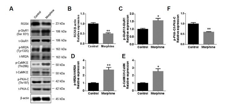
- Fig. 3 Morphine induced the activation of ionotropic glutamate receptors in primary NAc/striatal neurons To examine the effects of morphine on glutamate receptors in NAc/striatal neurons, the neurons (9 DIV) were incubated with morphine (10 muM) for 3 days. (A) Representative photographs of immunoblots for RGS4, phospho-GluR1 (Ser831), total-GluR1, phospho-NR2A (Tyr1325), total-NR2A, phospho-CaMKII (Thr286), total-CaMKII, phospho-PKA-C (Thr197), and total-PKA-C. (B-F) Bar graphs showing semi-quantitative analyses of the phosphorylation levels of GluR1, NR2A, CaMKII, and PKA-C in NAc/striatal neurons after vehicle and morphine treatment. All data are expressed as means +- SEM; n = 6 for each group and were analyzed with unpaired t-tests. p < 0.05 vs . vehicle-treated control and ** p < 0.01 vs. vehicle-treated control.