 Explore
 Validate
 Learn
Learn Western blot
 Immunohistochemistry
Antibody data
- Antibody Data
- Antigen structure
- References [3]
- Comments [0]
- Validations
- Immunohistochemistry [6]
Submit
Validation data
Reference
Comment
Report error
- Product number
- HPA007040 - Provider product page

- Provider
- Atlas Antibodies
- Proper citation
- Atlas Antibodies Cat#HPA007040, RRID:AB_1079185
- Product name
- Anti-KHK
- Antibody type
- Polyclonal
- Reactivity
- Human, Mouse, Rat
- Host
- Rabbit
- Conjugate
- Unconjugated
- Antigen sequence
YGDVVFVSKDVAKHLGFQSAEEALRGLYGRVRKGA
VLVCAWAEEGADALGPDGKLLHSDAFPPPRVVDTL
GAGDTFNASVIFSLSQGRSVQEALRFGCQV- Isotype
- IgG
- Vial size
- 100 µl
- Storage
- Store at +4°C for short term storage. Long time storage is recommended at -20°C.
Submitted references HIF-driven SF3B1 induces KHK-C to enforce fructolysis and heart disease.
Endogenous fructose production and metabolism in the liver contributes to the development of metabolic syndrome.
Opposing effects of fructokinase C and A isoforms on fructose-induced metabolic syndrome in mice
Mirtschink P, Krishnan J, Grimm F, Sarre A, Hörl M, Kayikci M, Fankhauser N, Christinat Y, Cortijo C, Feehan O, Vukolic A, Sossalla S, Stehr SN, Ule J, Zamboni N, Pedrazzini T, Krek W
Nature 2015 Jun 25;522(7557):444-449
Nature 2015 Jun 25;522(7557):444-449
Endogenous fructose production and metabolism in the liver contributes to the development of metabolic syndrome.
Lanaspa MA, Ishimoto T, Li N, Cicerchi C, Orlicky DJ, Ruzycki P, Rivard C, Inaba S, Roncal-Jimenez CA, Bales ES, Diggle CP, Asipu A, Petrash JM, Kosugi T, Maruyama S, Sanchez-Lozada LG, McManaman JL, Bonthron DT, Sautin YY, Johnson RJ
Nature communications 2013;4:2434
Nature communications 2013;4:2434
Opposing effects of fructokinase C and A isoforms on fructose-induced metabolic syndrome in mice
Ishimoto T, Lanaspa M, Le M, Garcia G, Diggle C, MacLean P, Jackman M, Asipu A, Roncal-Jimenez C, Kosugi T, Rivard C, Maruyama S, Rodriguez-Iturbe B, Sanchez-Lozada L, Bonthron D, Sautin Y, Johnson R
Proceedings of the National Academy of Sciences 2012 March;109(11):4320-4325
Proceedings of the National Academy of Sciences 2012 March;109(11):4320-4325
No comments: Submit comment
Enhanced validation
Supportive validation
- Submitted by
- Atlas Antibodies (provider)
- Enhanced method
- Orthogonal validation
- Main image

- Experimental details
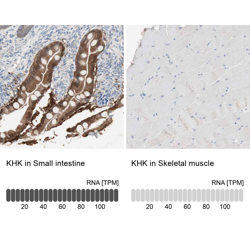
- Immunohistochemistry analysis in human small intestine and skeletal muscle tissues using HPA007040 antibody. Corresponding KHK RNA-seq data are presented for the same tissues.
- Sample type
- HUMAN
Supportive validation
- Submitted by
- Atlas Antibodies (provider)
- Main image

- Experimental details
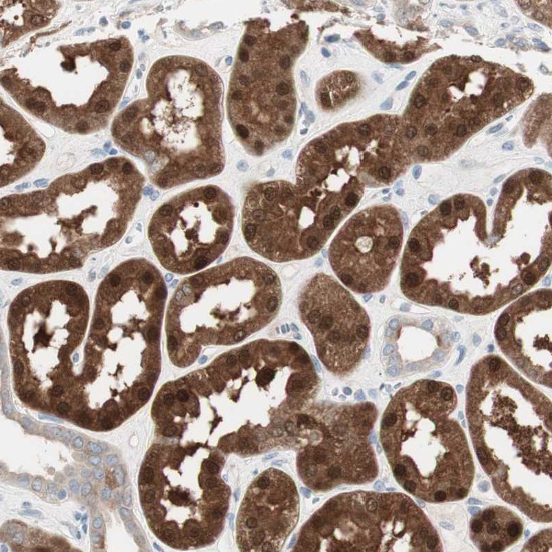
- Immunohistochemical staining of human kidney shows strong nuclear and cytoplasmic positivity in cells in tubules.
- Submitted by
- Atlas Antibodies (provider)
- Main image

- Experimental details
- Immunohistochemical staining of human small intestine shows moderate cytoplasmic positivity in glandular cells.
- Sample type
- HUMAN

- Submitted by
- Atlas Antibodies (provider)
- Main image
- Experimental details
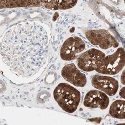
- Immunohistochemical staining of human kidney shows moderate cytoplasmic positivity in cells in tubules.
- Sample type
- HUMAN

- Submitted by
- Atlas Antibodies (provider)
- Main image

- Experimental details
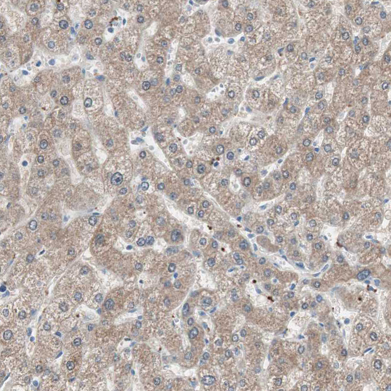
- Immunohistochemical staining of human liver shows moderate cytoplasmic positivity in hepatocytes.
- Sample type
- HUMAN
- Submitted by
- Atlas Antibodies (provider)
- Main image

- Experimental details
- Immunohistochemical staining of human skeletal muscle shows no positivity in myocytes as expected.
- Sample type
- HUMAN
