Explore
 Validate
 Learn
Learn Western blot
Antibody data
- Antibody Data
- Antigen structure
- References [1]
- Comments [0]
- Validations
- Western blot [3]
- Immunocytochemistry [1]
- Immunohistochemistry [1]
- Other assay [1]
Submit
Validation data
Reference
Comment
Report error
- Product number
- MA5-15675 - Provider product page

- Provider
- Invitrogen Antibodies
- Product name
- HK1 Monoclonal Antibody (7A7)
- Antibody type
- Monoclonal
- Antigen
- Purifed from natural sources
- Description
- MA5-15675 targets HK1 in IF, IHC, and WB applications and shows reactivity with Human, mouse, and Rat samples.
- Antibody clone number
- 7A7
- Concentration
- Conc. Not Determined
Submitted references Galactose protects against cell damage in mouse models of acute pancreatitis.
Peng S, Gerasimenko JV, Tsugorka TM, Gryshchenko O, Samarasinghe S, Petersen OH, Gerasimenko OV
The Journal of clinical investigation 2018 Aug 31;128(9):3769-3778
The Journal of clinical investigation 2018 Aug 31;128(9):3769-3778
No comments: Submit comment
Supportive validation
- Submitted by
- Invitrogen Antibodies (provider)
- Main image
- Experimental details
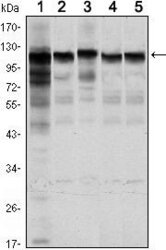
- Western blot analysis of HK1 using HK1 monoclonal antibody (Product # MA5-15675) in Jurkat (1), HeLa (2), HepG2 (3), MCF-7 (4) and PC-12 (5) cell lysate.

- Submitted by
- Invitrogen Antibodies (provider)
- Main image

- Experimental details
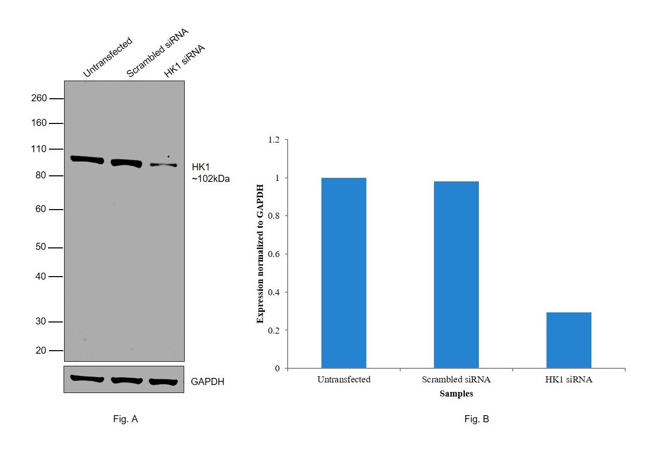
- Knockdown of HK1 was achieved by transfecting MCF7 with HK1 specific siRNAs (Silencer® select Product # S6559, S6558). Western blot analysis (Fig. a) was performed using Whole cell extracts from the HK1 knockdown cells (lane 3), non-targeting scrambled siRNA transfected cells (lane 2) and untransfected cells (lane 1). The blot was probed with HK1 Monoclonal Antibody (7A7) (Product # MA5-15675, 1/1000) and Goat anti-Mouse IgG (H+L) Superclonal™ Recombinant Secondary Antibody, HRP (Product # A28177, 1:4000). Densitometric analysis of this western blot is shown in histogram (Fig. b). Decrease in signal upon siRNA mediated knock down confirms that antibody is specific to HK1.
- Submitted by
- Invitrogen Antibodies (provider)
- Main image
- Experimental details
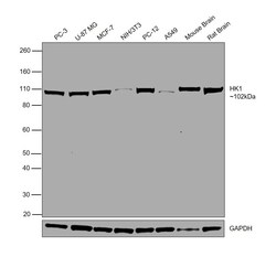
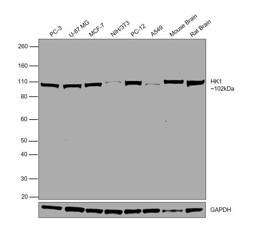
- Western blot was performed using Anti-HK1 Monoclonal Antibody (7A7)(Product # MA5-15675) and a 102 kDa band corresponding to HK1 was observed across all tested cell lines and tissues. Whole cell extracts (30 µg lysate) of PC-3 (Lane 1), U-87 MG (Lane 2), MCF7 (Lane 3), NIH/3T3 (Lane 4), PC-12 (Lane 5), A549 (Lane 6), Mouse Brain (Lane 7), Rat Brain (Lane 8) were electrophoresed using NuPAGE™ 4-12% Bis-Tris Protein Gel (Product # NP0322BOX). Resolved proteins were then transferred onto a Nitrocellulose membrane (Product # IB23001) by iBlot® 2 Dry Blotting System (Product # IB21001). The blot was probed with the primary antibody (1/1000) and detected by chemiluminescence with Goat anti-Mouse IgG (H+L) Superclonal™ Recombinant Secondary Antibody, HRP (Product # A28177,1:4000) using the iBright FL 1000 (Product # A32752). Chemiluminescent detection was performed using Novex® ECL Chemiluminescent Substrate Reagent Kit (Product # WP20005).
Supportive validation
- Submitted by
- Invitrogen Antibodies (provider)
- Main image
- Experimental details
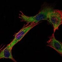
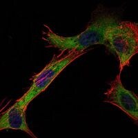
- Immunofluorescence analysis of NIH/3T3 cells using HK1 monoclonal antibody (Product # MA5-15675) (Green). Blue: DRAQ5 fluorescent DNA dye. Red: actin filaments have been labeled with phalloidin.
Supportive validation
- Submitted by
- Invitrogen Antibodies (provider)
- Main image
- Experimental details
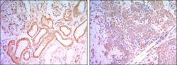
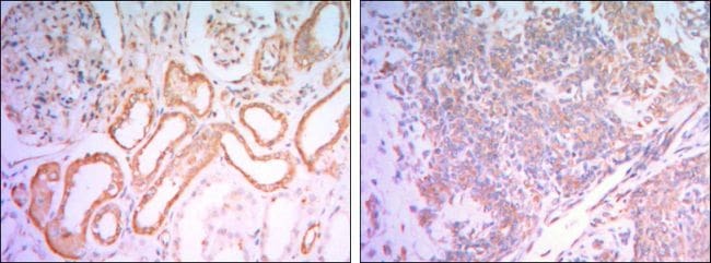
- Immunohistochemical analysis of paraffin-embedded human salivary gland tissues (left) and kidney tissues (right) using HK1 monoclonal antibody (Product # MA5-15675) followed with DAB staining.
Supportive validation
- Submitted by
- Invitrogen Antibodies (provider)
- Main image

- Experimental details
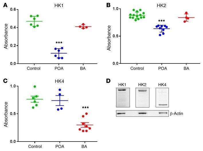
- Figure 6 HK activity is significantly inhibited in vitro by POA and BA. ( A ) HK1 activity is reduced significantly by 0.1 mM POA ( n = 6, P < 0.0001), but not changed significantly by 0.05% BA ( n = 4, P > 0.13) as compared with control ( n = 6). ( B ) HK2 activity is reduced significantly by 0.1 mM POA ( n = 9, P < 0.0001), but not affected by 0.05% BA ( n = 4, P > 0.3) as compared with control ( n = 13). ( C ) HK4 activity is reduced significantly by 0.05% BA ( n = 8, P < 0.0001), but not affected by 0.1 mM POA ( n = 4, P > 0.8) as compared with control ( n = 6). ( D ) Western blot analysis of the expression levels of HK1, HK2, and HK4 in PACs (representative case, repeated 3 times with similar results). *** P < 0.001, 1-way ANOVA.