Explore
 Validate
 Learn
LearnPA5-95382
antibody from Invitrogen Antibodies
Targeting: KDM5B
CT31, JARID1B, PLU-1, PPP1R98, RBBP2H1A
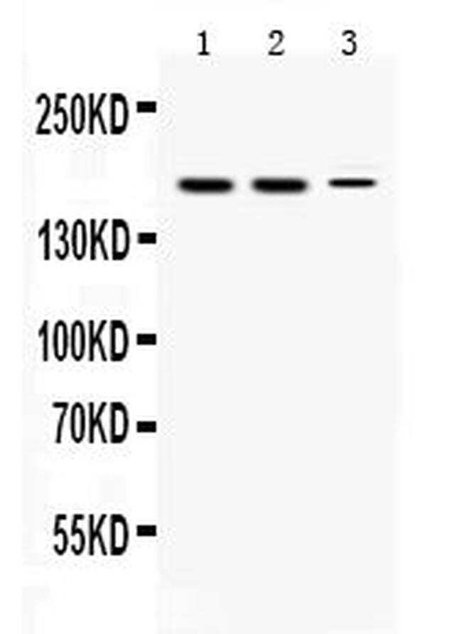
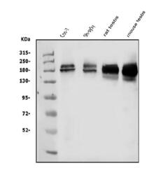
 Western blot
Antibody data
- Antibody Data
- Antigen structure
- References [0]
- Comments [0]
- Validations
- Western blot [2]
- Immunocytochemistry [1]
- Immunohistochemistry [6]
- Flow cytometry [1]
Submit
Validation data
Reference
Comment
Report error
- Product number
- PA5-95382 - Provider product page

- Provider
- Invitrogen Antibodies
- Product name
- KDM5B Polyclonal Antibody
- Antibody type
- Polyclonal
- Antigen
- Synthetic peptide
- Description
- Reconstitute with 0.2 mL of distilled water to yield a concentration of 500 µg/mL.
- Reactivity
- Human, Mouse, Rat
- Host
- Rabbit
- Isotype
- IgG
- Vial size
- 100 µg
- Concentration
- 500 µg/mL
- Storage
- Store at 4°C short term. For long term storage, store at -20°C, avoiding freeze/thaw cycles.
No comments: Submit comment
Supportive validation
- Submitted by
- Invitrogen Antibodies (provider)
- Main image
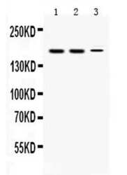
- Experimental details
- Western blot analysis of KDM5B in rat testis extract (lane 1), mouse testis extract (lane 2) and HEPG2 whole cell lysates (lane 3). Samples were incubated with KDM5B polyclonal antibody (Product # PA5-95382) using a 0.5 µg/mL dilution. developed was performed using enhanced chemiluminescence (ECL).
- Submitted by
- Invitrogen Antibodies (provider)
- Main image
- Experimental details
- Western blot analysis of KD.M5B in, Lane 1: monkey COS-7 whole cell lysates, Lane 2: human SH-SY5Y whole cell lysates, Lane 3: rat testis tissue lysates, Lane 4: mouse testis tissue lysates. Electrophoresis was performed on a 5-20% SDS-PAGE gel at 70V (Stacking gel) / 90V (Resolving gel) for 2-3 hours. The sample well of each lane was loaded with 50 µg of sample under reducing conditions. After Electrophoresis, proteins were transferred to a Nitrocellulose membrane at 150mA for 50-90 minutes. The membrane was blocked with 5% Non-fat Milk/ TBS for 1. 5 hour at RT. The membrane was incubated with KDM5B Polyclonal Antibody (Product # PA5-95382) at 0.5 μg/mL overnight at 4°C, then washed with TBS-0. 1% Tween 3 times with 5 minutes each and probed with a goat anti-rabbit IgG-HRP secondary antibody at a dilution of 1:10000 for 1. 5 hour at RT. The signal is developed using an Enhanced Chemiluminescent detection (ECL) kit. A specific band was detected for KD.M5B at approximately 180KD. The expected band size for KD.M5B is at 180KD.

Supportive validation
- Submitted by
- Invitrogen Antibodies (provider)
- Main image
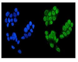
- Experimental details
- Immunocytochemistry analysis of KD.M5B using anti-KD.M5B antibody (Product # PA5-95382) . KD.M5B was detected in a section of MCF-7 cells. Enzyme antigen retrieval was performed using IHC enzyme antigen retrieval reagent for 15 mins. The cells were blocked with 10% goat serum and then incubated with 2μg/mL rabbit anti-KD.M5B antibody (Product # PA5-95382) overnight at 4°C. DyLight®488 Conjugated Goat Anti-Rabbit IgG was used as secondary antibody at 1:100 dilution and incubated for 30 minutes at 37°C. The section was counterstained with DAPI. Visualize using a fluorescence microscope and filter sets appropriate for the label used.

Supportive validation
- Submitted by
- Invitrogen Antibodies (provider)
- Main image
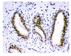
- Experimental details
- Immunohistochemical analysis of KD.M5B in paraffin-embedded section of human breast cancer tissue. Heat mediated antigen retrieval was performed in EDTA buffer (pH 8.0, epitope retrieval solution).The tissue section was blocked with 10% goat serum. The tissue section was then incubated with 1μg/mL rabbit anti-KD.M5B antibody (Product # PA5-95382) overnight at 4°C. Biotinylated goat anti-rabbit IgG was used as secondary antibody and incubated for 30 minutes at 37°C. The tissue section was developed using Strepavidin-Biotin-Complex (SABC) with DAB as the chromogen.
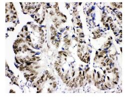
- Submitted by
- Invitrogen Antibodies (provider)
- Main image
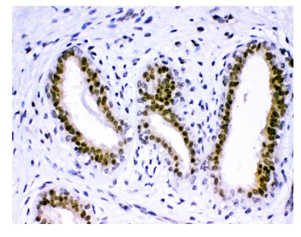
- Experimental details
- Immunohistochemical analysis of KD.M5B in paraffin-embedded section of human intestinal cancer tissue. Heat mediated antigen retrieval was performed in EDTA buffer (pH 8.0, epitope retrieval solution).The tissue section was blocked with 10% goat serum. The tissue section was then incubated with 1μg/mL rabbit anti-KD.M5B antibody (Product # PA5-95382) overnight at 4°C. Biotinylated goat anti-rabbit IgG was used as secondary antibody and incubated for 30 minutes at 37°C. The tissue section was developed using Strepavidin-Biotin-Complex (SABC) with DAB as the chromogen.
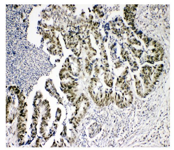
- Submitted by
- Invitrogen Antibodies (provider)
- Main image
- Experimental details
- Immunohistochemical analysis of KD.M5B in paraffin-embedded section of human intestinal cancer tissue. Heat mediated antigen retrieval was performed in EDTA buffer (pH 8.0, epitope retrieval solution).The tissue section was blocked with 10% goat serum. The tissue section was then incubated with 1μg/mL rabbit anti-KD.M5B antibody (Product # PA5-95382) overnight at 4°C. Biotinylated goat anti-rabbit IgG was used as secondary antibody and incubated for 30 minutes at 37°C. The tissue section was developed using Strepavidin-Biotin-Complex (SABC) with DAB as the chromogen.

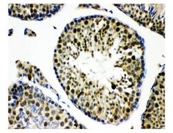
- Submitted by
- Invitrogen Antibodies (provider)
- Main image
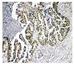
- Experimental details
- Immunohistochemical analysis of KD.M5B in paraffin-embedded section of human lung cancer tissue. Heat mediated antigen retrieval was performed in EDTA buffer (pH 8.0, epitope retrieval solution).The tissue section was blocked with 10% goat serum. The tissue section was then incubated with 1μg/mL rabbit anti-KD.M5B antibody (Product # PA5-95382) overnight at 4°C. Biotinylated goat anti-rabbit IgG was used as secondary antibody and incubated for 30 minutes at 37°C. The tissue section was developed using Strepavidin-Biotin-Complex (SABC) with DAB as the chromogen.

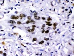
- Submitted by
- Invitrogen Antibodies (provider)
- Main image
- Experimental details
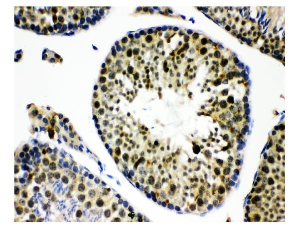
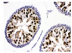
- Immunohistochemical analysis of KD.M5B in paraffin-embedded section of mouse testis tissue. Heat mediated antigen retrieval was performed in EDTA buffer (pH 8.0, epitope retrieval solution).The tissue section was blocked with 10% goat serum. The tissue section was then incubated with 1μg/mL rabbit anti-KD.M5B antibody (Product # PA5-95382) overnight at 4°C. Biotinylated goat anti-rabbit IgG was used as secondary antibody and incubated for 30 minutes at 37°C. The tissue section was developed using Strepavidin-Biotin-Complex (SABC) with DAB as the chromogen.
- Submitted by
- Invitrogen Antibodies (provider)
- Main image
- Experimental details
- Immunohistochemical analysis of KD.M5B in paraffin-embedded section of rat testis tissue. Heat mediated antigen retrieval was performed in EDTA buffer (pH 8.0, epitope retrieval solution).The tissue section was blocked with 10% goat serum. The tissue section was then incubated with 1μg/mL rabbit anti-KD.M5B antibody (Product # PA5-95382) overnight at 4°C. Biotinylated goat anti-rabbit IgG was used as secondary antibody and incubated for 30 minutes at 37°C. The tissue section was developed using Strepavidin-Biotin-Complex (SABC) with DAB as the chromogen.

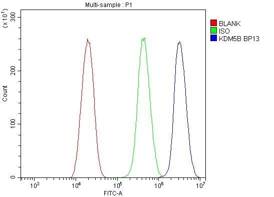
Supportive validation
- Submitted by
- Invitrogen Antibodies (provider)
- Main image
- Experimental details
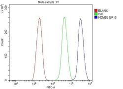
- Flow Cytometry of KDM5B in U2OS cells (blue line), isotype control rabbit IgG (green line) and unlabeled (red line). Samples were blocked with 10% goat serum, incubated with KDM5B Polyclonal Antibody (Product # PA5-95382) at a dilution of 1 μg (per 1x10^6 cells), followed by DyLight®488 conjugated goat anti-rabbit IgG (for 30 minutes at 20°C) using 5-10 μg (per 1x10^6 cells) dilution.