 Explore
 Validate
 Learn
Learn Western blot
Antibody data
- Antibody Data
- Antigen structure
- References [1]
- Comments [0]
- Validations
- Western blot [3]
- Immunocytochemistry [2]
- Immunohistochemistry [1]
- Other assay [1]
Submit
Validation data
Reference
Comment
Report error
- Product number
- PA5-29215 - Provider product page

- Provider
- Invitrogen Antibodies
- Product name
- RSK1 Polyclonal Antibody
- Antibody type
- Polyclonal
- Antigen
- Recombinant protein fragment
- Reactivity
- Human, Mouse
- Host
- Rabbit
- Isotype
- IgG
- Vial size
- 100 µL
- Concentration
- 1 mg/mL
- Storage
- Store at 4°C short term. For long term storage, store at -20°C, avoiding freeze/thaw cycles.
Submitted references The Preeclamptic Environment Promotes the Activation of Transcription Factor Kappa B by P53/RSK1 Complex in a HTR8/SVneo Trophoblastic Cell Line.
Sakowicz A, Bralewska M, Pietrucha T, Figueras F, Habrowska-Górczyńska DE, Piastowska-Ciesielska AW, Gach A, Sakowicz B, Rybak-Krzyszkowska M, Huras H, Grzesiak M, Biesiada L
International journal of molecular sciences 2021 Sep 22;22(19)
International journal of molecular sciences 2021 Sep 22;22(19)
No comments: Submit comment
Supportive validation
- Submitted by
- Invitrogen Antibodies (provider)
- Main image
- Experimental details
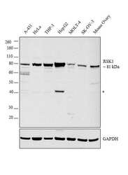
- Western blot analysis was performed on modified whole cell extract (1% SDS) (30 µg lysate) of A-431 (Lane 1), HeLa (Lane 2), THP-1 (Lane 3), Hep G2 (Lane 4), MOLT-4 (Lane 5), SK-OV-3 (Lane 6) and Mouse Ovary (Lane 7). The blot was probed with Anti-RSK1 Polyclonal Antibody (Product # PA5-29215, 1:5000 dilution) and detected by chemiluminescence using Goat anti-Rabbit IgG (H+L) Superclonal™ Secondary Antibody, HRP conjugate (Product # A27036, 0.25 µg/mL, 1:4000 dilution). A 81 kDa band corresponding to RSK1 was detected in all cell lines tested with a non-specific band (*) at ~40 kDa.

- Submitted by
- Invitrogen Antibodies (provider)
- Main image

- Experimental details
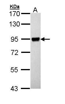
- Western Blot using RSK1 Polyclonal Antibody (Product # PA5-29215). Sample (30 µg of whole cell lysate). Lane A: Hep G2 . 7.5% SDS PAGE. RSK1 Polyclonal Antibody (Product # PA5-29215) diluted at 1:10,000.
- Submitted by
- Invitrogen Antibodies (provider)
- Main image
- Experimental details
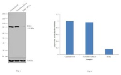
- Knockdown of RSK1 was achieved by transfecting HeLa with RSK1 specific siRNAs (Silencer® select Product # s12273; s12274). Western blot analysis (Fig. a) was performed using whole cell extracts from the RSK1 knockdown cells (lane 3), non-specific scrambled siRNA transfected cells (lane 2) and untransfected cells (lane 1). The blots were probed with RSK1 Polyclonal Antibody (Product # PA5-29215, 1:5000 dilution) and Goat anti-Rabbit IgG (H+L) Superclonal™ Secondary Antibody, HRP conjugate (Product # A27036, 0.25µg/mL, 1:4000 dilution). Densitometric analysis of this western blot is shown in histogram (Fig. b). Decrease in signal upon siRNA mediated knock down confirms that antibody is specific to RSK1.

Supportive validation
- Submitted by
- Invitrogen Antibodies (provider)
- Main image

- Experimental details
- Immunofluorescent analysis of RSK1 in methanol-fixed HeLa cells using a RSK1 polyclonal antibody (Product # PA5-29215) at a 1:200 dilution.

- Submitted by
- Invitrogen Antibodies (provider)
- Main image
- Experimental details
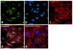
- Immunofluorescence analysis of RSK1 was performed using 70% confluent log phase HeLa cells. The cells were fixed with 4% paraformaldehyde for 10 minutes, permeabilized with 0.1% Triton™ X-100 for 15 minutes, and blocked with 1% BSA for 1 hour at room temperature. The cells were labeled with RSK1 Rabbit Polyclonal Antibody (Product # PA5-29215) at 1:200 dilution in 0.1% BSA, incubated at 4 degree Celsius overnight and then labeled with Goat anti-Rabbit IgG (H+L) Superclonal™ Secondary Antibody, Alexa Fluor® 488 conjugate (Product # A27034) at a dilution of 1:2000 for 45 minutes at room temperature (Panel a: green). Nuclei (Panel b: blue) were stained with ProLong™ Diamond Antifade Mountant with DAPI (Product # P36962). F-actin (Panel c: red) was stained with Rhodamine Phalloidin (Product # R415). Panel d represents the merged image showing Nuclear as well as Cytoplasmic localization. Panel e represents control cells with no primary antibody to assess background. The images were captured at 60X magnification.

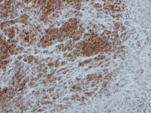
Supportive validation
- Submitted by
- Invitrogen Antibodies (provider)
- Main image
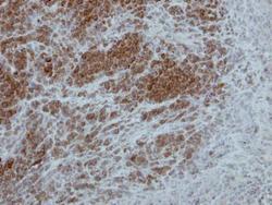
- Experimental details
- Immunohistochemical analysis of paraffin-embedded SCM1 xenograft, using p90 RSK1 (Product # PA5-29215) antibody at 1:500 dilution. Antigen Retrieval: Citrate buffer, pH 6.0, 15 min.
Supportive validation
- Submitted by
- Invitrogen Antibodies (provider)
- Main image

- Experimental details
- Figure 3 Duolink II PLA assay results for HTR8/SVneo cells cultured in normoxic and hypoxic conditions in medium containing 1% serum from preeclamptic ( b , d ) or normotensive ( a , c ) women. Each p53/RSK1 complex in HTR8/SVneo cells is visualised as an individual fluorescence red spot, nuclei were labelled with DAPI. Red spot - complex of p53/RSK1; blue spot - nucleus of cell. Magnification 40x; scale bar 50 um.
