Explore
 Validate
 Learn
Learn Western blot
 Flow cytometry
Antibody data
- Antibody Data
- Antigen structure
- References [1]
- Comments [0]
- Validations
- Western blot [5]
- Immunocytochemistry [2]
- Immunohistochemistry [1]
Submit
Validation data
Reference
Comment
Report error
- Product number
- PA5-27720 - Provider product page

- Provider
- Invitrogen Antibodies
- Product name
- Calpain 2 Polyclonal Antibody
- Antibody type
- Polyclonal
- Antigen
- Recombinant protein fragment
- Description
- The recommended shelf life for this product is 1 year from date of receipt.
- Reactivity
- Human, Mouse
- Host
- Rabbit
- Isotype
- IgG
- Vial size
- 100 µL
- Concentration
- 1 mg/mL
- Storage
- Store at 4°C short term. For long term storage, store at -20°C, avoiding freeze/thaw cycles.
Submitted references Comparison of the protein expression of calpain-1, calpain-2, calpastatin and calmodulin between gastric cancer and normal gastric mucosa.
Liu B, Zhou Y, Lu D, Liu Y, Zhang SQ, Xu Y, Li W, Gu X
Oncology letters 2017 Sep;14(3):3705-3710
Oncology letters 2017 Sep;14(3):3705-3710
No comments: Submit comment
Supportive validation
- Submitted by
- Invitrogen Antibodies (provider)
- Main image

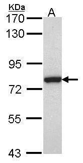
- Experimental details
- Western blot analysis of Calpain 2 using 30 µg of Jurkat lysate. Samples were loaded onto a 7.5% SDS-PAGE gel and probed with a Calpain 2 polyclonal antibody (Product # PA5-27720) at a dilution of 1:1000.
- Submitted by
- Invitrogen Antibodies (provider)
- Main image
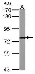
- Experimental details
- Western blot analysis of Calpain 2 using 30 µg of NIH-3T3 lysate. Samples were loaded onto a 7.5% SDS-PAGE gel and probed with a Calpain 2 polyclonal antibody (Product # PA5-27720) at a dilution of 1:1000.

- Submitted by
- Invitrogen Antibodies (provider)
- Main image

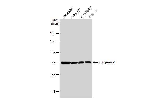
- Experimental details
- Western Blot using Calpain 2 Polyclonal Antibody (Product # PA5-27720). Various whole cell extracts (30 µg) were separated by 7.5% SDS-PAGE, and the membrane was blotted with Calpain 2 Polyclonal Antibody (Product # PA5-27720) diluted at 1:3,000. The HRP-conjugated anti-rabbit IgG antibody was used to detect the primary antibody.
- Submitted by
- Invitrogen Antibodies (provider)
- Main image

- Experimental details
- Knockdown of Calpain 2 (CAPN2) was achieved by transfecting A-431 with Calpain 2 (CAPN2) specific siRNAs (Silencer® select Products # s321). Western blot analysis (Fig. a) was performed using Whole cell extracts from the Calpain 2 (CAPN2) knockdown cells (Lane 3), non-specific scrambled siRNA transfected cells (Lane 2) and untransfected cells (Lane 1). The blot was probed with Calpain 2 Polyclonal Antibody (Product # PA5-27720, 1:1000 dilution) and Goat anti-Rabbit IgG (H+L) Superclonal™ Recombinant Secondary Antibody, HRP (Product # A27036, 1:4000 dilution). Densitometric analysis of this western blot is shown in histogram (Fig. b). Reduction of signal upon siRNA mediated knock down confirms that antibody is specific to CAPN2.

- Submitted by
- Invitrogen Antibodies (provider)
- Main image

- Experimental details
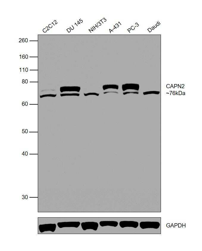
- Western blot was performed using Anti-Calpain 2 Polyclonal Antibody (Product # PA5-27720) and a 76 kDa band corresponding to Calpain 2 was observed in C2C12, DU 145, A-431 and PC-3, but not in NIH/3T3 and Daudi. Whole cell lysates (30 µg lysate) of C2C12 (Lane 1), DU 145 (Lane 2), NIH/3T3 (Lane 3), A-431 (Lane 4), PC-3 (Lane 5) and Daudi (Lane 6) were electrophoresed using NuPAGE® 10 % Bis-Tris gel (Product # NP0302BOX). Resolved proteins were then transferred onto a nitrocellulose membrane (Product # IB23001) by iBlot® 2 Dry Blotting System (Product # IB21001). The blots were probed with the primary antibody (1:1000 dilution) and detected by chemiluminescence with Goat anti-Rabbit IgG (H+L), Superclonal™ Recombinant Secondary Antibody, HRP (Product # A27036, 1:4000 dilution) using the iBright FL 1000 (Product # A32752). Chemiluminescent detection was performed using Novex® ECL Chemiluminescent Substrate Reagent Kit (Product # WP20005). An uncharacterized band was observed at ~70 kDa.
Supportive validation
- Submitted by
- Invitrogen Antibodies (provider)
- Main image
- Experimental details
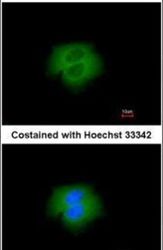
- Immunofluorescent analysis of Calpain 2 in paraformaldehyde-fixed A431 cells using a Calpain 2 polyclonal antibody (Product # PA5-27720) at a 1:200 dilution.

- Submitted by
- Invitrogen Antibodies (provider)
- Main image
- Experimental details
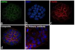
- Immunofluorescence analysis of CAPN2 was performed using A-431 cells. The cells were fixed with 4% paraformaldehyde for 10 minutes, permeabilized with 0.1% Triton™ X-100 for 15 minutes, and blocked with 2% BSA for 1 hour at room temperature. The cells were labeled with Calpain 2 Polyclonal Antibody (Product # PA5-27720) at 5 µg/mL in 0.1% BSA, incubated at 4 degree Celsius overnight and then labeled with Goat anti-Rabbit IgG (H+L) Superclonal™ Recombinant Secondary Antibody, Alexa Fluor® 488 (Product # A27034) at a dilution of 1:2000 for 45 minutes at room temperature (Panel a: green). Nuclei (Panel b: blue) were stained with ProLong™ Diamond Antifade Mountant with DAPI (Product # P36962). F-actin (Panel c: red) was stained with Rhodamine Phalloidin (Product # R415, 1:300). Panel d represents the merged image showing cytoplasmic localization. Panel e represents control cells with no primary antibody to assess background. The images were captured at 60X magnification.

Supportive validation
- Submitted by
- Invitrogen Antibodies (provider)
- Main image

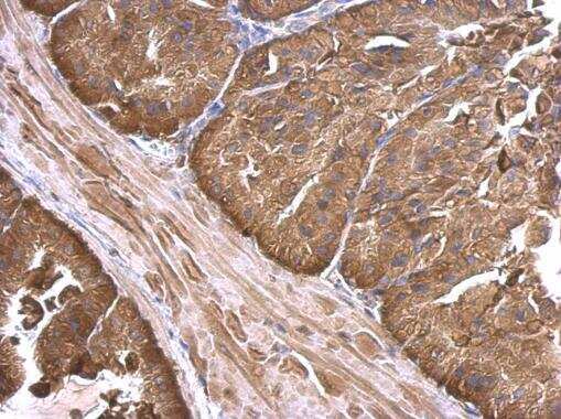
- Experimental details
- Calpain 2 Polyclonal Antibody detects Calpain 2 protein at cytosol on mouse prostate by immunohistochemical analysis. Sample: Paraffin-embedded mouse prostate. Calpain 2 Polyclonal Antibody (Product # PA5-27720) dilution: 1:500. Antigen Retrieval: EDTA based buffer, pH 8.0, 15 min.