Explore
 Validate
 Learn
Learn Western blot
Antibody data
- Antibody Data
- Antigen structure
- References [0]
- Comments [0]
- Validations
- Western blot [5]
- Immunocytochemistry [2]
- Immunohistochemistry [1]
Submit
Validation data
Reference
Comment
Report error
- Product number
- PA5-78291 - Provider product page

- Provider
- Invitrogen Antibodies
- Product name
- ROCK2 Polyclonal Antibody
- Antibody type
- Polyclonal
- Antigen
- Synthetic peptide
- Description
- Positive Control: 293T, A431, HeLa, HepG2
- Concentration
- 0.192 mg/mL
No comments: Submit comment
Supportive validation
- Submitted by
- Invitrogen Antibodies (provider)
- Main image
- Experimental details
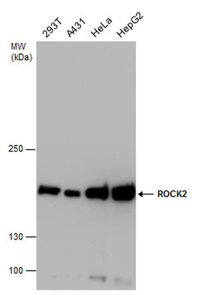
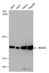
- Western blot analysis of ROCK2 in whole cell lysate using 30 µg of protein. Samples were separated with 5% SDS-PAGE and incubated with ROCK2 polyclonal antibody (Product # PA5-78291) using a dilution of 1:5000.
- Submitted by
- Invitrogen Antibodies (provider)
- Main image
- Experimental details
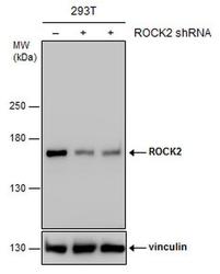
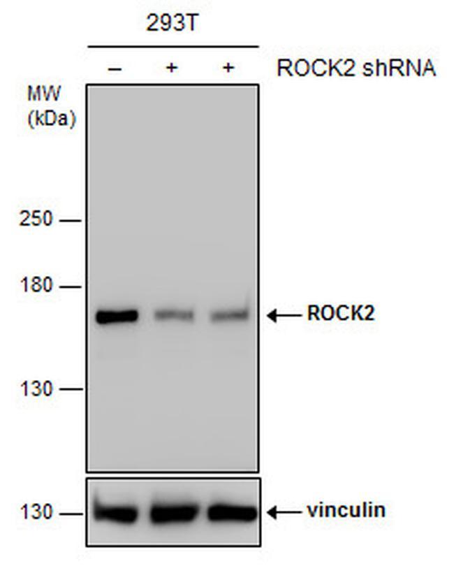
- Western blot analysis of ROCK2 in non-transfected (-) and transfected (+) 293T cells using 30 µg of protein. Samples were separated with 5% SDS-PAGE and incubated with ROCK2 polyclonal antibody (Product # PA5-78291) using a dilution of 1:4000.
- Submitted by
- Invitrogen Antibodies (provider)
- Main image
- Experimental details
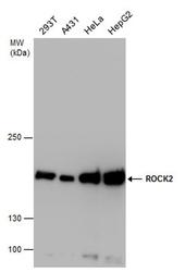
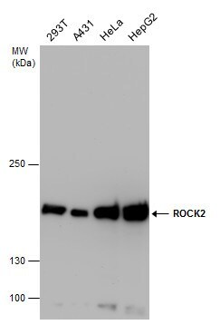
- ROCK2 Polyclonal Antibody detects ROCK2 protein by western blot analysis. Various whole cell extracts (30 µg) were separated by 5% SDS-PAGE, and the membrane was blotted with ROCK2 Polyclonal Antibody (Product # PA5-78291) diluted at a dilution of 1:5,000.
- Submitted by
- Invitrogen Antibodies (provider)
- Main image
- Experimental details
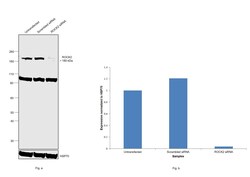
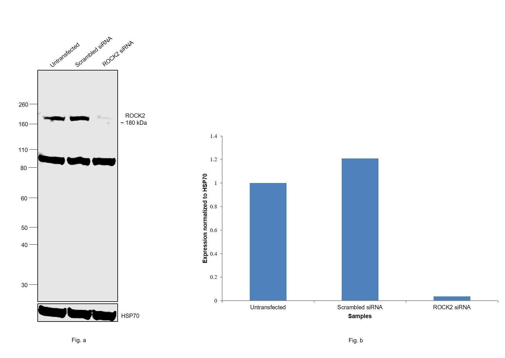
- Knockdown of ROCK2 was achieved by transfecting HeLa with ROCK2 specific siRNAs (Silencer® select Product # s18162). Western blot analysis (Fig. a) was performed using whole cell extracts from the ROCK2 knockdown cells (lane 3), non-specific scrambled siRNA transfected cells (lane 2) and untransfected cells (lane 1). The blot was probed with ROCK2 Polyclonal Antibody (Product # PA5-78291, 1:5000 dilution) and Goat anti-Rabbit IgG (H+L) Superclonal™ Recombinant Secondary Antibody, HRP (Product # A27036, 1:4000 dilution). Densitometric analysis of this western blot is shown in histogram (Fig. b). Decrease in signal upon siRNA mediated knock down confirms that antibody is specific to ROCK2. An uncharacterized band around 90 kDa was also observed.
- Submitted by
- Invitrogen Antibodies (provider)
- Main image
- Experimental details
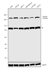
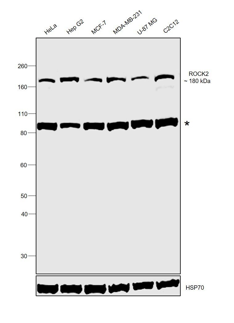
- Western blot was performed using Anti-ROCK2 Polyclonal Antibody (Product # PA5-78291) and a 180 kDa corresponding to ROCK2 along with an uncharacterized band around 90 kDa was observed across cell lines tested. Whole cell extracts (30 µg lysate) of HeLa (Lane 1), Hep G2 (Lane 2), MCF-7 (Lane 3), MDA-MB-231 (Lane 4), U-87 MG (Lane 5) and C1C12 (Lane 6) were electrophoresed using NuPAGE™ 4-12% Bis-Tris Protein Gel (Product # NP0322BOX). Resolved proteins were then transferred onto a nitrocellulose membrane (Product # IB23001) by XCell SureLock™ Mini-Cell and XCell II™ Blot Module (Product # EI0002). The blot was probed with the primary antibody (1:5000 dilution) and detected by chemiluminescence with Goat anti-Rabbit IgG (H+L) Superclonal™ Recombinant Secondary Antibody, HRP (Product # A27036, 1:4000 dilution) using the iBright FL 1000 (Product # A32752). Chemiluminescent detection was performed using Novex® ECL Chemiluminescent Substrate Reagent Kit (Product # WP20005).
Supportive validation
- Submitted by
- Invitrogen Antibodies (provider)
- Main image

- Experimental details
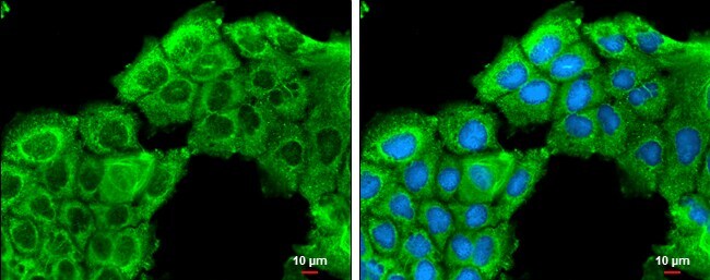
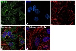
- ROCK2 Polyclonal Antibody detects ROCK2 protein at cytoplasm by immunofluorescent analysis. Sample: A431 cells were fixed in ice-cold MeOH for 5 min. Green: ROCK2 protein stained by ROCK2 Polyclonal Antibody (Product # PA5-78291) diluted at 1:500. Blue: Hoechst 33342 staining. Scale bar = 10 µm.
- Submitted by
- Invitrogen Antibodies (provider)
- Main image
- Experimental details
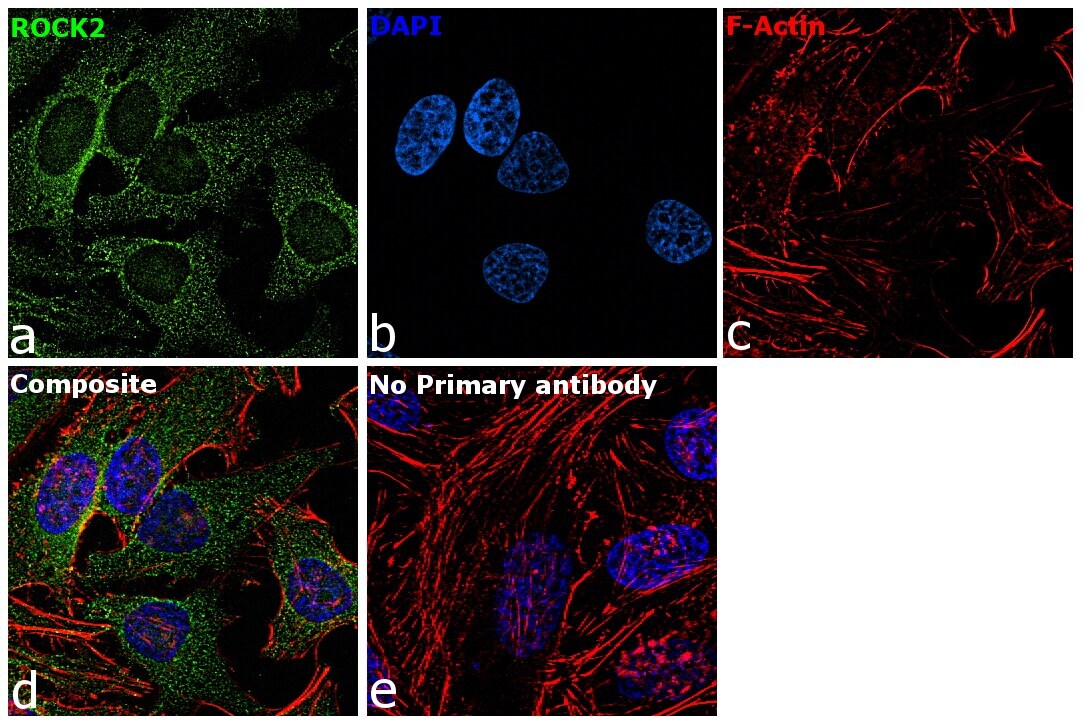
- Immunofluorescence analysis of ROCK2 was performed using 70% confluent log phase HeLa cells. The cells were fixed with 4% paraformaldehyde for 10 minutes, permeabilized with 0.1% Triton™ X-100 for 15 minutes, and blocked with 2% BSA for 1 hour at room temperature. The cells were labeled with ROCK2 Rabbit Polyclonal Antibody (Product # PA5-78291) at 5 µg/mL in 0.1% BSA, incubated at 4 degree Celsius overnight and then with Goat anti-Rabbit IgG (H+L) Highly Cross-Adsorbed Secondary Antibody, Alexa Fluor Plus 488 (Product # A32731) at a dilution of 1:2000 for 45 minutes at room temperature (Panel a: green). Nuclei (Panel b: blue) were stained with ProLong™ Diamond Antifade Mountant with DAPI (Product # P36962). F-actin (Panel c: red) was stained with Rhodamine Phalloidin (Product # R415, 1:300). Panel d represents the merged image showing cytoplasmic localization. Panel e represents control cells with no primary antibody to assess background. The images were captured at 60X magnification.
Supportive validation
- Submitted by
- Invitrogen Antibodies (provider)
- Main image
- Experimental details
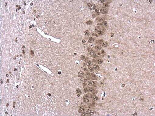
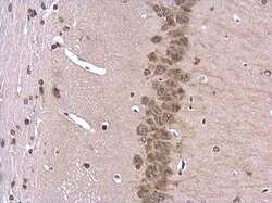
- ROCK2 Polyclonal Antibody detects ROCK2 protein at cytoplasm and nucleus in mouse brain by immunohistochemical analysis. Sample: Paraffin-embedded mouse brain. ROCK2 Polyclonal Antibody (Product # PA5-78291) diluted at 1:500. Antigen Retrieval: Citrate buffer, pH 6.0, 15 min.