 Explore
 Validate
 Learn
Learn Western blot
Antibody data
- Antibody Data
- Antigen structure
- References [0]
- Comments [0]
- Validations
- Western blot [2]
- Immunohistochemistry [1]
Submit
Validation data
Reference
Comment
Report error
- Product number
- GTX16685 - Provider product page

- Provider
- GeneTex
- Product name
- KCNK1 antibody
- Antibody type
- Polyclonal
- Reactivity
- Human, Mouse, Rat
- Host
- Rabbit
No comments: Submit comment
Supportive validation
- Submitted by
- GeneTex (provider)
- Main image

- Experimental details
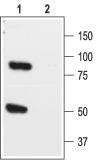
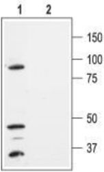
- Anti-K2P1.1_(TWIK-1)_(extracellular) - Western blot analysis of rat brain lysate: 1. Anti-K2P1.1 (TWIK-1) (extracellular) antibody, (1:200). 2. Anti-K2P1.1 (TWIK-1) (extracellular) antibody, preincubated with the control peptide antigen.
- Submitted by
- GeneTex (provider)
- Main image
- Experimental details
- Anti-K2P1.1_(TWIK-1)_(extracellular) - Western blot analysis of HEK-293-K2P1.1 transfected cells: 1. Anti-K2P1.1 (TWIK-1) (extracellular) antibody, (1:300). 2. Anti-K2P1.1 (TWIK-1) (extracellular) antibody, preincubated with the control peptide antigen.

Supportive validation
- Submitted by
- GeneTex (provider)
- Main image

- Experimental details
- Anti-K2P1.1_(TWIK-1)_(extracellular) - Expression of K2P1.1 in mouse cerebellum Immunohistochemical staining of mouse cerebellum using Anti-K2P1.1 (TWIK-1) (extracellular) antibody. A. K2P1.1 channel appears in glial processes (red) . B. staining of Purkinje nerve cells with mouse anti calbindin D28K (a calcium binding protein, green). C. Confocal merge of K2P1.1 channel and calbindin D28K demonstrates the separate localization of these proteins.
