 Explore
 Validate
 Learn
Learn Western blot
Antibody data
- Antibody Data
- Antigen structure
- References [3]
- Comments [0]
- Validations
- Western blot [1]
- Immunohistochemistry [1]
Submit
Validation data
Reference
Comment
Report error
- Product number
- MAB397-100 - Provider product page

- Provider
- R&D Systems
- Product name
- Human/Mouse/Rat TrkB Antibody
- Antibody type
- Monoclonal
- Description
- Protein A or G purified from hybridoma culture supernatant. Detects human TrkB in direct ELISAs. Detects human, mouse, and rat TrkB in Western blots. In ELISAs and Western blots, no cross-reactivity with recombinant human (rh) TrkA, rhTrkC, or recombinant rat TrkA is observed.
- Reactivity
- Human, Mouse, Rat
- Host
- Mouse
- Conjugate
- Unconjugated
- Antigen sequence
Q16620- Isotype
- IgG
- Antibody clone number
- 75133
- Vial size
- 100 ug
- Storage
- Use a manual defrost freezer and avoid repeated freeze-thaw cycles. 12 months from date of receipt, -20 to -70 °C as supplied. 1 month, 2 to 8 °C under sterile conditions after reconstitution. 6 months, -20 to -70 °C under sterile conditions after reconstitution.
Submitted references Brain-derived neurotrophic factor (BDNF)-induced tropomyosin-related kinase B (Trk B) signaling is a potential therapeutic target for peritoneal carcinomatosis arising from colorectal cancer.
Brain-derived neurotrophic factor is increased in serum and skin levels of patients with chronic spontaneous urticaria.
IL-10 inhibits endothelium-dependent T cell costimulation by up-regulation of ILT3/4 in human vascular endothelial cells.
Tanaka K, Okugawa Y, Toiyama Y, Inoue Y, Saigusa S, Kawamura M, Araki T, Uchida K, Mohri Y, Kusunoki M
PloS one 2014;9(5):e96410
PloS one 2014;9(5):e96410
Brain-derived neurotrophic factor is increased in serum and skin levels of patients with chronic spontaneous urticaria.
Rössing K, Novak N, Mommert S, Pfab F, Gehring M, Wedi B, Kapp A, Raap U
Clinical and experimental allergy : journal of the British Society for Allergy and Clinical Immunology 2011 Oct;41(10):1392-9
Clinical and experimental allergy : journal of the British Society for Allergy and Clinical Immunology 2011 Oct;41(10):1392-9
IL-10 inhibits endothelium-dependent T cell costimulation by up-regulation of ILT3/4 in human vascular endothelial cells.
Gleissner CA, Zastrow A, Klingenberg R, Kluger MS, Konstandin M, Celik S, Haemmerling S, Shankar V, Giese T, Katus HA, Dengler TJ
European journal of immunology 2007 Jan;37(1):177-92
European journal of immunology 2007 Jan;37(1):177-92
No comments: Submit comment
Supportive validation
- Submitted by
- R&D Systems (provider)
- Main image
- Experimental details
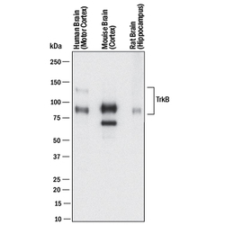
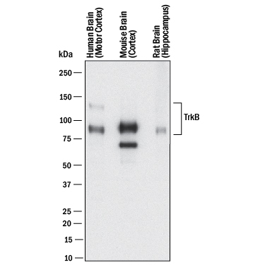
- Detection of Human, Mouse, and Rat TrkB by Western Blot. Western blot shows lysates of human brain (motor cortex) tissue, mouse brain (cortex) tissue, and rat brain (hippocampus) tissue. PVDF membrane was probed with 2 µg/mL of Mouse Anti-Human/Mouse/Rat TrkB Monoclonal Antibody (Catalog # MAB397) followed by HRP-conjugated Anti-Mouse IgG Secondary Antibody (Catalog # HAF018). Specific bands were detected for TrkB at approximately 95 kDa and 145 kDa (as indicated). This experiment was conducted under reducing conditions and using Immunoblot Buffer Group 1.
Supportive validation
- Submitted by
- R&D Systems (provider)
- Main image
- Experimental details
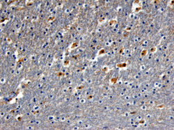
- TrkB in Human Brain. TrkB was detected in immersion fixed paraffin-embedded sections of human brain (hippocampus) using Mouse Anti-Human/Mouse/Rat TrkB Monoclonal Antibody (Catalog # MAB397) at 25 µg/mL overnight at 4 °C. Before incubation with the primary antibody tissue was subjected to heat-induced epitope retrieval using Antigen Retrieval Reagent-Basic (Catalog # CTS013). Tissue was stained using the Anti-Mouse HRP-DAB Cell & Tissue Staining Kit (brown; Catalog # CTS002) and counterstained with hematoxylin (blue). View our protocol for Chromogenic IHC Staining of Paraffin-embedded Tissue Sections.
