 Explore
 Validate
 Learn
Learn Western blot
Antibody data
- Antibody Data
- Antigen structure
- References [0]
- Comments [0]
- Validations
- Western blot [1]
- Immunohistochemistry [2]
Submit
Validation data
Reference
Comment
Report error
- Product number
- AF1519 - Provider product page

- Provider
- R&D Systems
- Product name
- Human p38 delta Antibody
- Antibody type
- Polyclonal
- Description
- Antigen Affinity-purified. Detects endogenous human p38 delta . Mouse and rat p38 delta reactivity has not been demonstrated but is likely due to their high homology with human p38 delta . This antibody does not detect recombinant p38 alpha , p38 beta or p38 gamma in Western blots.
- Reactivity
- Human
- Host
- Rabbit
- Conjugate
- Unconjugated
- Antigen sequence
O15264- Isotype
- IgG
- Vial size
- 100 ug
- Concentration
- LYOPH
- Storage
- Use a manual defrost freezer and avoid repeated freeze-thaw cycles. 12 months from date of receipt, -20 to -70 °C as supplied. 1 month, 2 to 8 °C under sterile conditions after reconstitution. 6 months, -20 to -70 °C under sterile conditions after reconstitution.
No comments: Submit comment
Supportive validation
- Submitted by
- R&D Systems (provider)
- Main image

- Experimental details
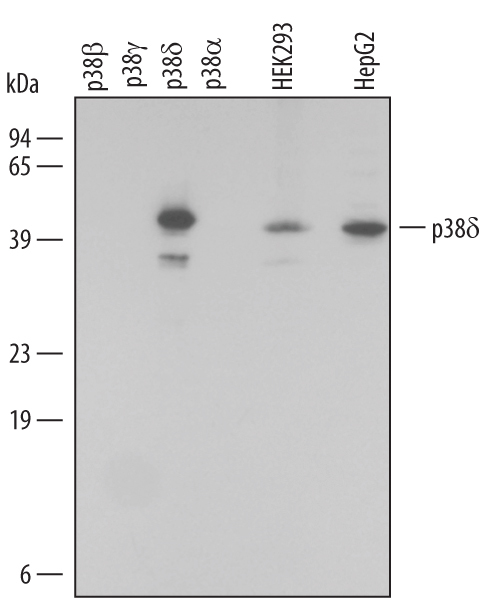
- Detection of Human p38 delta by Western Blot. Western blot shows lysates of HEK293 human embryonic kidney cell line and HepG2 human hepatocellular carcinoma cell line. PVDF membrane was probed with 0.1 µg/mL Rabbit Anti-Human p38 delta Antigen Affinity-purified Polyclonal Antibody (Catalog # AF1519) followed by HRP-conjugated Anti-Rabbit IgG Secondary Antibody (Catalog # HAF008). For additional reference, recombinant p38 beta , p38 gamma , p38 delta , and p38 alpha at 2 ng/lane) were included. A specific band for p38 delta was detected at approximately 42 kDa (as indicated). This experiment was conducted under reducing conditions and using Immunoblot Buffer Group 1.
Supportive validation
- Submitted by
- R&D Systems (provider)
- Main image

- Experimental details
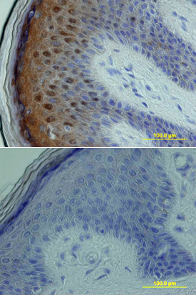
- p38 delta in Human Skin. p38 delta was detected in immersion fixed paraffin-embedded sections of human skin using Rabbit Anti-Human p38 delta Antigen Affinity-purified Polyclonal Antibody (Catalog # AF1519) at 15 µg/mL overnight at 4 °C. Tissue was stained using the Anti-Rabbit HRP-DAB Cell & Tissue Staining Kit (brown; Catalog # CTS005) and counterstained with hematoxylin (blue). Lower panel shows a lack of labeling if primary antibodies are omitted and tissue is stained only with secondary antibody followed by incubation with detection reagents. View our protocol for Chromogenic IHC Staining of Paraffin-embedded Tissue Sections.
- Submitted by
- R&D Systems (provider)
- Main image
- Experimental details
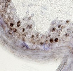
- p38 delta in Human Skin. p38 delta was detected in immersion fixed paraffin-embedded sections of human skin using 15 µg/mL Rabbit Anti-Human p38 delta Antigen Affinity-purified Polyclonal Antibody (Catalog # AF1519) overnight at 4 °C. Tissue was stained with the Anti-Rabbit HRP-DAB Cell & Tissue Staining Kit (brown; Catalog # CTS005) and counterstained with hematoxylin (blue). View our protocol for Chromogenic IHC Staining of Paraffin-embedded Tissue Sections.
