Antibody data
- Antibody Data
- Antigen structure
- References [7]
- Comments [0]
- Validations
- Western blot [2]
- Immunohistochemistry [11]
- Other assay [2]
Submit
Validation data
Reference
Comment
Report error
- Product number
- UM800118 - Provider product page

- Provider
- OriGene
- Product name
- ALK mouse monoclonal antibody, clone OTI1A4
- Antibody type
- Monoclonal
- Description
- ALK mouse monoclonal antibody, clone OTI1A4
- Host
- Mouse
- Conjugate
- Unconjugated
- Epitope
- ALK
- Isotype
- IgG
- Antibody clone number
- OTI1A4
- Vial size
- 100 µl
- Concentration
- 1 mg/ml
Submitted references Antibody 1A4 with routine immunohistochemistry demonstrates high sensitivity for ALK rearrangement screening of Chinese lung adenocarcinoma patients: A single-center large-scale study.
[Statement of the German Society for Pathology and the working group thoracic oncology of the working group oncology/German Cancer Society on ALK testing in NSCLC: Immunohistochemistry and/or FISH?].
Reliability Assurance of Detection of EML4-ALK Rearrangement in Non-Small Cell Lung Cancer: The Results of Proficiency Testing in China.
[ALK-Diagnostics in NSCLC - Immunohistochemistry (IHC) and/or Fluorescence-in-situ Hybridisation (FISH)].
Inflammatory myofibroblastic tumour of the bladder in children: a review.
A novel, highly sensitive ALK antibody 1A4 facilitates effective screening for ALK rearrangements in lung adenocarcinomas by standard immunohistochemistry.
Extraordinary response to crizotinib in a woman with squamous cell lung cancer after two courses of failed chemotherapy.
Wang Q, Zhao L, Yang X, Wei S, Zeng Y, Mao C, Lin L, Fu P, Lyu L, Li Z, Xiao H
Lung cancer (Amsterdam, Netherlands) 2016 May;95:39-43
Lung cancer (Amsterdam, Netherlands) 2016 May;95:39-43
[Statement of the German Society for Pathology and the working group thoracic oncology of the working group oncology/German Cancer Society on ALK testing in NSCLC: Immunohistochemistry and/or FISH?].
von Laffert M, Schirmacher P, Warth A, Weichert W, Büttner R, Huber RM, Wolf J, Griesinger F, Dietel M, Grohé C
Der Pathologe 2016 Mar;37(2):187-91
Der Pathologe 2016 Mar;37(2):187-91
Reliability Assurance of Detection of EML4-ALK Rearrangement in Non-Small Cell Lung Cancer: The Results of Proficiency Testing in China.
Li Y, Zhang R, Peng R, Ding J, Han Y, Wang G, Zhang K, Lin G, Li J
Journal of thoracic oncology : official publication of the International Association for the Study of Lung Cancer 2016 Jun;11(6):924-9
Journal of thoracic oncology : official publication of the International Association for the Study of Lung Cancer 2016 Jun;11(6):924-9
[ALK-Diagnostics in NSCLC - Immunohistochemistry (IHC) and/or Fluorescence-in-situ Hybridisation (FISH)].
von Laffert M, Schirmacher P, Warth A, Weichert W, Büttner R, Huber RM, Wolf J, Griesinger F, Dietel M, Grohé Ch
Pneumologie (Stuttgart, Germany) 2016 Apr;70(4):277-81
Pneumologie (Stuttgart, Germany) 2016 Apr;70(4):277-81
Inflammatory myofibroblastic tumour of the bladder in children: a review.
Collin M, Charles A, Barker A, Khosa J, Samnakay N
Journal of pediatric urology 2015 Oct;11(5):239-45
Journal of pediatric urology 2015 Oct;11(5):239-45
A novel, highly sensitive ALK antibody 1A4 facilitates effective screening for ALK rearrangements in lung adenocarcinomas by standard immunohistochemistry.
Gruber K, Kohlhäufl M, Friedel G, Ott G, Kalla C
Journal of thoracic oncology : official publication of the International Association for the Study of Lung Cancer 2015 Apr;10(4):713-6
Journal of thoracic oncology : official publication of the International Association for the Study of Lung Cancer 2015 Apr;10(4):713-6
Extraordinary response to crizotinib in a woman with squamous cell lung cancer after two courses of failed chemotherapy.
Wang Q, He Y, Yang X, Wang Y, Xiao H
BMC pulmonary medicine 2014 May 15;14:83
BMC pulmonary medicine 2014 May 15;14:83
No comments: Submit comment
Supportive validation
- Submitted by
- OriGene (provider)
- Main image
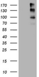
- Experimental details
- HEK293T cells were transfected with the pCMV6-ENTRY control (Left lane) or pCMV6-ENTRY ALK (RC222485, Right lane) cDNA for 48 hrs and lysed. Equivalent amounts of cell lysates (5 ug per lane) were separated by SDS-PAGE and immunoblotted with anti-ALK.
- Validation comment
- WB
- Submitted by
- OriGene (provider)
- Main image
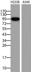
- Experimental details
- Western blot analysis of extracts (35ug) from H2228 and A549 cell lines by using anti-ALK monoclonal antibody. (UM800118, 1:10,000)
- Validation comment
- WB
Supportive validation
- Submitted by
- OriGene (provider)
- Main image
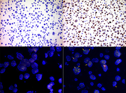
- Experimental details
- Immunohistochemistry staining of paraffin-embedded human cell line H1975 (upper left) and H2228 (upper right) on IHC antibody quality control slide using anti-ALK mouse monoclonal antibody UM800118 (1:400). The ALK rearrangement in H2228 cells is labeled with ALK Breakapart probe in FISH test (lower right, 60X) and the control of H1975 cell at the same FISH probe test (lower left, 60X).
- Validation comment
- IHC
- Submitted by
- OriGene (provider)
- Main image
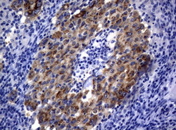
- Experimental details
- Immunohistochemical staining of paraffin-embedded Human non-small cell lung cancer sample with EML4-ALK translocation detected by PCR using anti-ALK mouse monoclonal antibody. (UM800118, 1:50; heat-induced epitope retrieval by 1mM EDTA in 10mM Tris, pH8.0, 120C for 3min)
- Validation comment
- IHC
- Submitted by
- OriGene (provider)
- Main image
- Experimental details
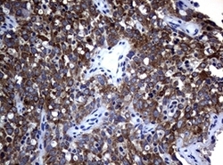
- Immunohistochemical staining of paraffin-embedded Human non-small cell lung cancer sample with ALK translocation detected by FISH using anti-ALK mouse monoclonal antibody. (UM800118, 1:50; heat-induced epitope retrieval by 1mM EDTA in 10mM Tris, pH8.0, 120C for 3min)
- Validation comment
- IHC
- Submitted by
- OriGene (provider)
- Main image
- Experimental details
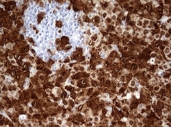
- Immunohistochemical staining of paraffin-embedded Human large B cell lymphoma with ALK translocation using anti-ALK mouse monoclonal antibody. (UM800118, 1:50; heat-induced epitope retrieval by 1mM EDTA in 10mM Tris, pH8.0, 120C for 3min)
- Validation comment
- IHC
- Submitted by
- OriGene (provider)
- Main image
- Experimental details
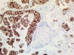
- Immunohistochemical staining of paraffin-embedded human ALK-positive lung cancer tissue using anti-ALK mouse monoclonal antibody. (UM800118, 1:100 for 30 min at RT; heat-induced epitope retrieval by TEE, pH9.0)
- Validation comment
- IHC
- Submitted by
- OriGene (provider)
- Main image
- Experimental details
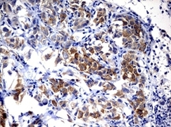
- Immunohistochemical staining of paraffin-embedded ALK-positive lung tumor xenograft using anti-ALK mouse monoclonal antibody. (UM800118, 1:50; heat-induced epitope retrieval by 1mM EDTA in 10mM Tris, pH8.0, 120C for 3min)
- Validation comment
- IHC
- Submitted by
- OriGene (provider)
- Main image
- Experimental details
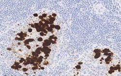
- Figure from citation: Optimal ALK staining of the ALCL with ALK rearrangement using the mAb clone OTI1A4 optimally calibrated, HIER in TRS High pH 9 (Dako), a 3-step polymer based detection system and performed on Omnis, Dako.
- Validation comment
- IHC
- Submitted by
- OriGene (provider)
- Main image
- Experimental details
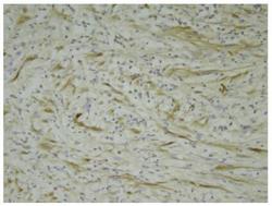
- Figure from citation: Immunohistochemistry showing brown Anaplastic lymphoma kinase (ALK-1) staining of spindle cells in an inflammatory myofibroblastic tumour of the bladder specimen.
- Validation comment
- IHC
- Submitted by
- OriGene (provider)
- Main image
- Experimental details
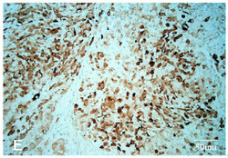
- Figure from citation: Immunohistochemistry of ALK protein level by using anti-ALK antibody in human right cervical lymph node. Dilution: 1:200
- Validation comment
- IHC
- Submitted by
- OriGene (provider)
- Main image

- Experimental details
- Figure from citation: ALK staining in human adenocarcinoma specimens using antibodies 1A4 and D5F3. Case #119 (ALK-negative according to qRT-PCR) is clearly negative with D5F3 (right) but weakly positive with 1A4 (1+) (left).
- Validation comment
- IHC
- Submitted by
- OriGene (provider)
- Main image
- Experimental details
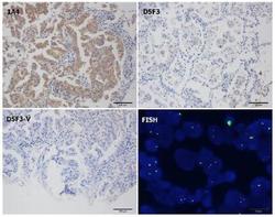
- Figure from citation: ALK rearrangements detected by 1A4 with routine IHC, D5F3 with routine IHC, and D5F3 with Ventana system and FISH assay in tissues of consecutive patients with lung adenocarcinoma. 1A4 (+), D5F3 (-), D5F3 Ventana (-), and FISH (+).
- Validation comment
- IHC
Supportive validation
- Submitted by
- OriGene (provider)
- Main image
- Experimental details
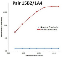
- ALK Luminex with 15B2 Capture (TA801288) and 1A4 Detection (UM800118) Antibodies. Substrate used: full length HEK293 cells expressed recombinant ALK protein (TP322485).
- Validation comment
- LMNX
- Submitted by
- OriGene (provider)
- Main image

- Experimental details
- ALK Luminex with 10D7 Capture (TA801306) and 1A4 Detection (UM800118) Antibodies. Substrate used: full length HEK293 cells expressed recombinant ALK protein (TP322485).
- Validation comment
- LMNX
 Explore
 Validate
 Learn
Learn Western blot