Explore
 Validate
 Learn
Learn Western blot
 Immunocytochemistry
Antibody data
- Antibody Data
- Antigen structure
- References [2]
- Comments [0]
- Validations
- Western blot [5]
- Immunoprecipitation [1]
- Immunohistochemistry [2]
Submit
Validation data
Reference
Comment
Report error
- Product number
- NB500-186 - Provider product page

- Provider
- Novus Biologicals
- Proper citation
- Novus Cat#NB500-186, RRID:AB_10002798
- Product name
- Rabbit Polyclonal CCAR1 Antibody
- Antibody type
- Polyclonal
- Description
- Immunogen affinity purified.
- Reactivity
- Human, Mouse
- Host
- Rabbit
- Isotype
- IgG
- Vial size
- 100 ul
- Concentration
- 1.0 mg/ml
- Storage
- Store at 4C. Do not freeze.
Submitted references Necdin enhances myoblasts survival by facilitating the degradation of the mediator of apoptosis CCAR1/CARP1.
Requirement of cell cycle and apoptosis regulator 1 for target gene activation by Wnt and beta-catenin and for anchorage-independent growth of human colon carcinoma cells.
François S, D'Orlando C, Fatone T, Touvier T, Pessina P, Meneveri R, Brunelli S
PloS one 2012;7(8):e43335
PloS one 2012;7(8):e43335
Requirement of cell cycle and apoptosis regulator 1 for target gene activation by Wnt and beta-catenin and for anchorage-independent growth of human colon carcinoma cells.
Ou CY, Kim JH, Yang CK, Stallcup MR
The Journal of biological chemistry 2009 Jul 31;284(31):20629-37
The Journal of biological chemistry 2009 Jul 31;284(31):20629-37
No comments: Submit comment
Supportive validation
- Submitted by
- Novus Biologicals (provider)
- Main image
- Experimental details
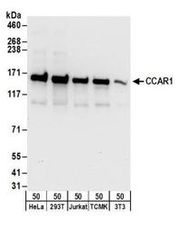
- Western Blot: CCAR1 Antibody [NB500-186] - Detection of Human and Mouse CCAR1 by Western Blot. Samples: Whole cell lysate (50 ug) from HeLa, 293T, Jurkat, mouse TCMK-1, and mouse NIH3T3 cells. Antibodies: Affinity purified rabbit anti-CCAR1 antibody NB500-186 used for WB at 0.1 ug/ml. Detection: Chemiluminescence with an exposure time of 10 seconds.

- Submitted by
- Novus Biologicals (provider)
- Main image

- Experimental details
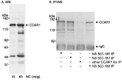
- Western Blot: CCAR1 Antibody [NB500-186] - Detection of Human CCAR1on HeLa whole cell lysate using NB500-186. CCAR1 was also IPed with another rabbit anti-CCAR1 antibody, NB500-187, and NB500-188. IPed CCAR1 was blotted using NB500-188.
- Submitted by
- Novus Biologicals (provider)
- Main image

- Experimental details
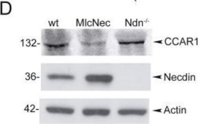
- Western Blot: CCAR1 Antibody [NB500-186] - Necdin controls CCAR1 protein abundance in myogenic cells. Representative western blot showing expression of CCAR1 and necdin in primary myoblasts isolated from wt, Ndn-/-, tgMlcNec newborn mice. Proteins were detected using antibodies specific for CCAR1, necdin and beta-actin as control. Image collected and cropped by CiteAb from the following publication (http://dx.plos.org/10.1371/journal.pone.0043335), licensed under a CC-BY licence.
- Submitted by
- Novus Biologicals (provider)
- Main image
- Experimental details
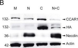
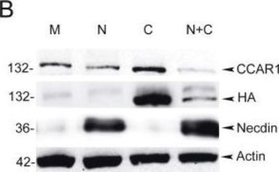
- Western Blot: CCAR1 Antibody [NB500-186] - Necdin controls CCAR1 protein abundance in myogenic cells. Representative western blot showing CCAR1 and necdin (beta-actin as loading control) expression in C2C12 mock transfected (M) or transfected with pSG5-HA-CCAR1 and/or pCMV-FLAG-Necdin (N or N+C). Both the endogenous and transfected CCAR1 were detected with the polyclonal anti-CCAR1, the transfected CCAR1 with anti-HA. Image collected and cropped by CiteAb from the following publication (http://dx.plos.org/10.1371/journal.pone.0043335), licensed under a CC-BY licence.
- Submitted by
- Novus Biologicals (provider)
- Main image
- Experimental details
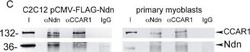
- Western Blot: CCAR1 Antibody [NB500-186] - Necdin interacts with CCAR1/CARP1. Interaction of necdin and CCAR1 in C2C12 myoblasts and primary myoblast cells. Co-IP experiments were performed on protein extract from C2C12 transfected with pCMV-FLAG-Necdin (C2C12 pCMV-FLAG-Ndn) or primary myoblasts from C57/Bl6 newborn mice. Proteins were immunoprecipitated using the polyclonal anti-Ndn (alpha-Ndn) or polyclonal anti-CCAR1 (alpha-CCAR1) and with a non-specific rabbit antisera as control (IgG). Necdin and CCAR1 were detected in immunoprecipitated samples using specific antibody. Input (I) represents 10% of the immunoprecipitated proteins. Image collected and cropped by CiteAb from the following publication (http://dx.plos.org/10.1371/journal.pone.0043335), licensed under a CC-BY licence.

Supportive validation
- Submitted by
- Novus Biologicals (provider)
- Main image

- Experimental details
- Immunoprecipitation: CCAR1 Antibody [NB500-186] - Detection of human CCAR1 by western blot of immunoprecipitates. Samples: Whole cell lysate (1 mg for IP; 20% of IP loaded) from HeLa cells. Antibodies: Affinity purified rabbit anti-CCAR1 antibody NB500-186 (lot NB500-186-2) used for IP at 6 ug/mg lysate. CCAR1 was also immunoprecipitated by a previous lot (lot NB500-186-1) of the antibody. For blotting immunoprecipitated CCAR1, NB500-186 was used at 1 ug/ml. Detection: Chemiluminescence with an exposure time of 10 seconds.

Supportive validation
- Submitted by
- Novus Biologicals (provider)
- Main image
- Experimental details
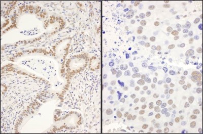
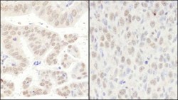
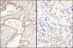
- Immunohistochemistry-Paraffin: CCAR1 Antibody [NB500-186] - Human stomach carcinoma (left) and mouse squamous cell carcinoma (right). Antibody used at a dilution of 1:1000 (1ug/ml).

- Submitted by
- Novus Biologicals (provider)
- Main image
- Experimental details
- Immunohistochemistry-Paraffin: CCAR1 Antibody [NB500-186] - Human stomach carcinoma (left) and mouse renal cell carcinoma (right). Antibody: Affinity purified rabbit anti-CCAR1 used at a dilution of 1:1,000 (1ug/ml). Detection: DAB