Explore
 Validate
 Learn
Learn Western blot
Antibody data
- Antibody Data
- Antigen structure
- References [0]
- Comments [0]
- Validations
- Western blot [2]
- Immunocytochemistry [1]
- Flow cytometry [1]
- Other assay [1]
Submit
Validation data
Reference
Comment
Report error
- Product number
- MA5-29406 - Provider product page

- Provider
- Invitrogen Antibodies
- Product name
- Latexin Recombinant Rabbit Monoclonal Antibody (101)
- Antibody type
- Monoclonal
- Antigen
- Recombinant full-length protein
- Description
- This product is preservative free. It is recommended to add sodium azide to avoid contamination (final concentration 0.05%-0.1%).
- Antibody clone number
- 101
- Concentration
- 1 mg/mL
No comments: Submit comment
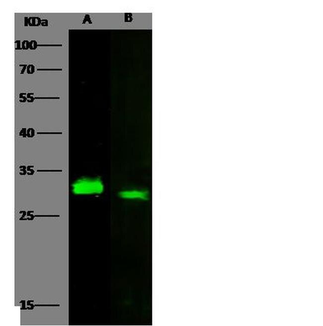
Supportive validation
- Submitted by
- Invitrogen Antibodies (provider)
- Main image

- Experimental details
- Western blot analysis of Latexin in Lane A: A549 Whole Cell Lysate (30 µg). Samples were probed using a Latexin Monoclonal Antibody (Product # MA5-29406) at a 1:500 dilution, followed by a Goat Anti-Rabbit IgG (H+L), Dylight 800 Secondary Antibody at a 1:10000 dilution. Western blot was performed under reducing conditions. Predicted band size:26 kDa. Observed band size:26 kDa.
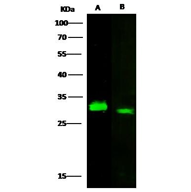
- Submitted by
- Invitrogen Antibodies (provider)
- Main image

- Experimental details
- Western Blot using Latexin Recombinant Rabbit Monoclonal Antibody (101) (Product # MA5-29406) at 1:500 dilution. Lane A: A549 Whole Cell Lysate. Lysates/proteins at 30 μg per lane. Secondary Goat Anti-Rabbit IgG H&L (DyLight™ 800) at 1:10,000 dilution. Developed using the Odyssey technique. Performed under reducing conditions. Predicted band size: 26 kDa. Observed band size: 26 kDa.
Supportive validation
- Submitted by
- Invitrogen Antibodies (provider)
- Main image

- Experimental details
- Immunofluorescence staining of Human Latexin in MCF7 cells. Cells were fixed with 4% PFA, permeabilzed with 1% Triton X-100 in PBS, blocked with 10% serum, and incubated with Latexin Recombinant Rabbit Monoclonal Antibody (101) (Product # MA5-29406, 1:60) at 4°C overnight. Then cells were stained with the Alexa Fluor® 488-conjugated Goat Anti-rabbit IgG secondary antibody (green) and counterstained with DAPI (blue). Positive staining was localized to cytoplasm.

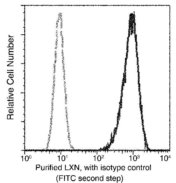
Supportive validation
- Submitted by
- Invitrogen Antibodies (provider)
- Main image

- Experimental details
- Flow cytometric analysis of Human Latexin expression on MCF-7 cells. The cells were treated according to manufacturer’s manual, stained with Latexin Recombinant Rabbit Monoclonal Antibody (101) (Product # MA5-29406), then a FITC-conjugated Secondary antibody. The fluorescence histograms were derived from gated events with the forward and side light-scatter characteristics of intact cells.
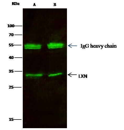
Supportive validation
- Submitted by
- Invitrogen Antibodies (provider)
- Main image

- Experimental details
- Latexin Immunoprecipitation using: Lane A: 0.5 mg A549 Whole Cell Lysate, Lane B: 0.5 mg Hela Whole Cell Lysate 2 µL with Latexin Recombinant Rabbit Monoclonal Antibody (101) (Product # MA5-29406) and 15 µL of 50 % Protein G agarose. Primary antibody: Latexin Recombinant Rabbit Monoclonal Antibody (101), at 1:200 dilution. Secondary antibody: Dylight 800-labeled antibody to rabbit IgG (H+L), at 1:5,000 dilution. Developed using the Odyssey technique. Performed under reducing conditions. Predicted band size: 25.7 kDa. Observed band size: 25.7 kDa.