 Explore
 Validate
 Learn
Learn Western blot
 Immunohistochemistry
Antibody data
- Antibody Data
- Antigen structure
- References [0]
- Comments [0]
- Validations
- Immunohistochemistry [23]
Submit
Validation data
Reference
Comment
Report error
- Product number
- LS-C662555 - Provider product page

- Provider
- LSBio
- Product name
- PEBP1 / RKIP Antibody LS-C662555
- Antibody type
- Polyclonal
- Reactivity
- Human
- Host
- Rabbit
- Storage
- At -20°C for 1 year. After reconstitution, at 4°C for 1 month. It can also be aliquotted and stored frozen at -20°C for a longer time. Avoid freeze-thaw cycles.
No comments: Submit comment
Supportive validation
- Submitted by
- LSBio (provider)
- Enhanced method
- Genetic validation
- Main image

- Experimental details
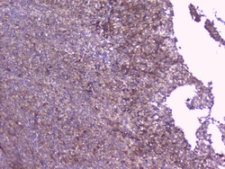
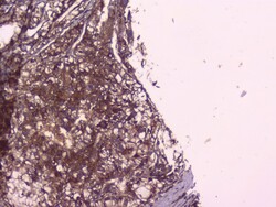
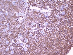
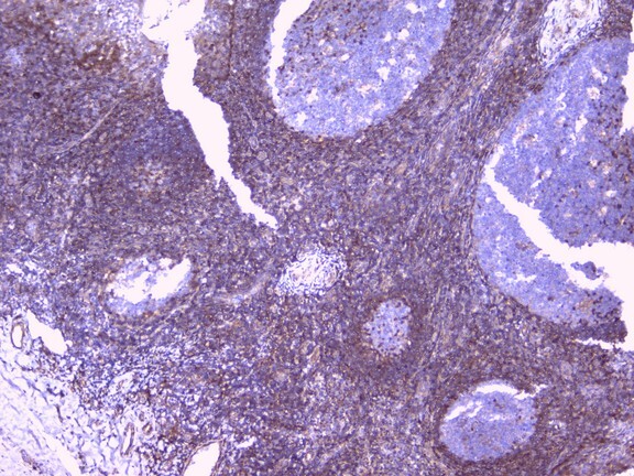
- IHC analysis of PBP using anti-PBP antibody. PBP was detected in paraffin-embedded section of human intestinal cancer tissue. Heat mediated antigen retrieval was performed in citrate buffer (pH6, epitope retrieval solution) for 20 mins. The tissue section was blocked with 10% goat serum. The tissue section was then incubated with 1µg/ml rabbit anti-PBP Antibody overnight at 4°C. Biotinylated goat anti-rabbit IgG was used as secondary antibody and incubated for 30 minutes at 37°C. The tissue section was developed using Strepavidin-Biotin-Complex (SABC) with DAB as the chromogen.

- Submitted by
- LSBio (provider)
- Enhanced method
- Genetic validation
- Main image
- Experimental details
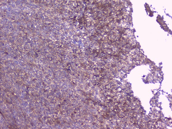
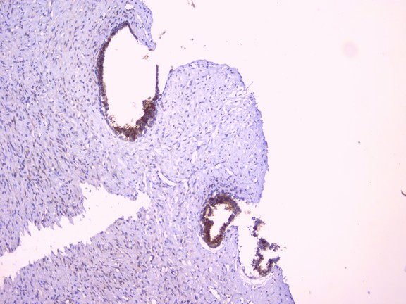
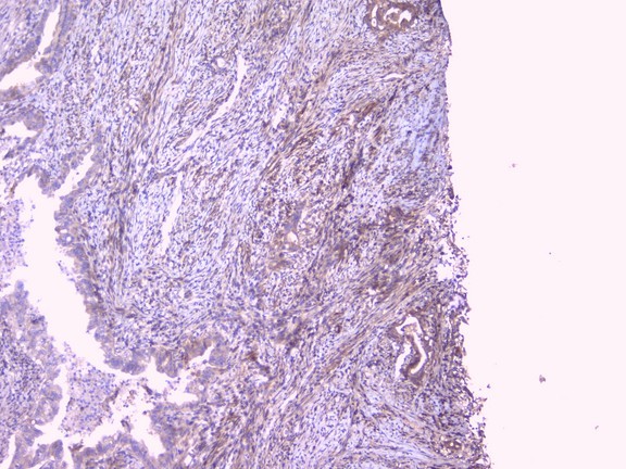
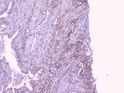
- IHC analysis of PBP using anti-PBP antibody. PBP was detected in paraffin-embedded section of human sarcoma tissue. Heat mediated antigen retrieval was performed in citrate buffer (pH6, epitope retrieval solution) for 20 mins. The tissue section was blocked with 10% goat serum. The tissue section was then incubated with 1µg/ml rabbit anti-PBP Antibody overnight at 4°C. Biotinylated goat anti-rabbit IgG was used as secondary antibody and incubated for 30 minutes at 37°C. The tissue section was developed using Strepavidin-Biotin-Complex (SABC) with DAB as the chromogen.
- Submitted by
- LSBio (provider)
- Enhanced method
- Genetic validation
- Main image
- Experimental details
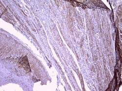
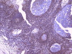
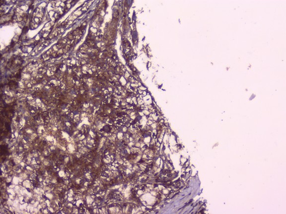
- IHC analysis of PBP using anti-PBP antibody. PBP was detected in paraffin-embedded section of human endometrial carcinoma tissue. Heat mediated antigen retrieval was performed in citrate buffer (pH6, epitope retrieval solution) for 20 mins. The tissue section was blocked with 10% goat serum. The tissue section was then incubated with 1µg/ml rabbit anti-PBP Antibody overnight at 4°C. Biotinylated goat anti-rabbit IgG was used as secondary antibody and incubated for 30 minutes at 37°C. The tissue section was developed using Strepavidin-Biotin-Complex (SABC) with DAB as the chromogen.
- Submitted by
- LSBio (provider)
- Enhanced method
- Genetic validation
- Main image
- Experimental details
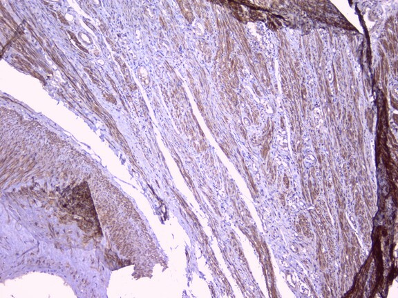
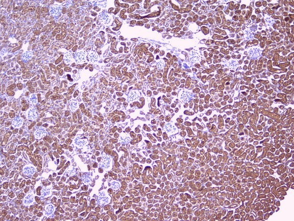
- IHC analysis of PBP using anti-PBP antibody. PBP was detected in paraffin-embedded section of human liver cancer tissue. Heat mediated antigen retrieval was performed in citrate buffer (pH6, epitope retrieval solution) for 20 mins. The tissue section was blocked with 10% goat serum. The tissue section was then incubated with 1µg/ml rabbit anti-PBP Antibody overnight at 4°C. Biotinylated goat anti-rabbit IgG was used as secondary antibody and incubated for 30 minutes at 37°C. The tissue section was developed using Strepavidin-Biotin-Complex (SABC) with DAB as the chromogen.

- Submitted by
- LSBio (provider)
- Enhanced method
- Genetic validation
- Main image

- Experimental details
- IHC analysis of PBP using anti-PBP antibody. PBP was detected in paraffin-embedded section of human mammary cancer tissue. Heat mediated antigen retrieval was performed in citrate buffer (pH6, epitope retrieval solution) for 20 mins. The tissue section was blocked with 10% goat serum. The tissue section was then incubated with 1µg/ml rabbit anti-PBP Antibody overnight at 4°C. Biotinylated goat anti-rabbit IgG was used as secondary antibody and incubated for 30 minutes at 37°C. The tissue section was developed using Strepavidin-Biotin-Complex (SABC) with DAB as the chromogen.
- Submitted by
- LSBio (provider)
- Enhanced method
- Genetic validation
- Main image

- Experimental details
- IHC analysis of PBP using anti-PBP antibody. PBP was detected in paraffin-embedded section of human pancreatic cancer tissue. Heat mediated antigen retrieval was performed in citrate buffer (pH6, epitope retrieval solution) for 20 mins. The tissue section was blocked with 10% goat serum. The tissue section was then incubated with 1µg/ml rabbit anti-PBP Antibody overnight at 4°C. Biotinylated goat anti-rabbit IgG was used as secondary antibody and incubated for 30 minutes at 37°C. The tissue section was developed using Strepavidin-Biotin-Complex (SABC) with DAB as the chromogen.

- Submitted by
- LSBio (provider)
- Enhanced method
- Genetic validation
- Main image
- Experimental details
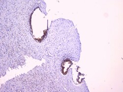
- IHC analysis of PBP using anti-PBP antibody. PBP was detected in paraffin-embedded section of human tonsil tissue. Heat mediated antigen retrieval was performed in citrate buffer (pH6, epitope retrieval solution) for 20 mins. The tissue section was blocked with 10% goat serum. The tissue section was then incubated with 1µg/ml rabbit anti-PBP Antibody overnight at 4°C. Biotinylated goat anti-rabbit IgG was used as secondary antibody and incubated for 30 minutes at 37°C. The tissue section was developed using Strepavidin-Biotin-Complex (SABC) with DAB as the chromogen.

- Submitted by
- LSBio (provider)
- Enhanced method
- Genetic validation
- Main image

- Experimental details
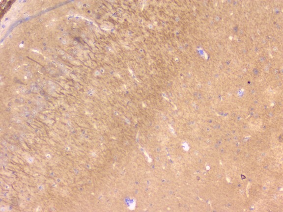
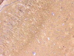
- IHC analysis of PBP using anti-PBP antibody. PBP was detected in paraffin-embedded section of mouse brain tissue. Heat mediated antigen retrieval was performed in citrate buffer (pH6, epitope retrieval solution) for 20 mins. The tissue section was blocked with 10% goat serum. The tissue section was then incubated with 1µg/ml rabbit anti-PBP Antibody overnight at 4°C. Biotinylated goat anti-rabbit IgG was used as secondary antibody and incubated for 30 minutes at 37°C. The tissue section was developed using Strepavidin-Biotin-Complex (SABC) with DAB as the chromogen.
- Submitted by
- LSBio (provider)
- Enhanced method
- Genetic validation
- Main image
- Experimental details
- IHC analysis of PBP using anti-PBP antibody. PBP was detected in paraffin-embedded section of mouse kidney tissue. Heat mediated antigen retrieval was performed in citrate buffer (pH6, epitope retrieval solution) for 20 mins. The tissue section was blocked with 10% goat serum. The tissue section was then incubated with 1µg/ml rabbit anti-PBP Antibody overnight at 4°C. Biotinylated goat anti-rabbit IgG was used as secondary antibody and incubated for 30 minutes at 37°C. The tissue section was developed using Strepavidin-Biotin-Complex (SABC) with DAB as the chromogen.
- Submitted by
- LSBio (provider)
- Main image

- Experimental details
- IHC analysis of PBP using anti-PBP antibody. PBP was detected in paraffin-embedded section of mouse kidney tissue. Heat mediated antigen retrieval was performed in citrate buffer (pH6, epitope retrieval solution) for 20 mins. The tissue section was blocked with 10% goat serum. The tissue section was then incubated with 1µg/ml rabbit anti-PBP Antibody overnight at 4°C. Biotinylated goat anti-rabbit IgG was used as secondary antibody and incubated for 30 minutes at 37°C. The tissue section was developed using Strepavidin-Biotin-Complex (SABC) with DAB as the chromogen.

- Submitted by
- LSBio (provider)
- Main image

- Experimental details
- IHC analysis of PBP using anti-PBP antibody. PBP was detected in paraffin-embedded section of human intestinal cancer tissue. Heat mediated antigen retrieval was performed in citrate buffer (pH6, epitope retrieval solution) for 20 mins. The tissue section was blocked with 10% goat serum. The tissue section was then incubated with 1µg/ml rabbit anti-PBP Antibody overnight at 4°C. Biotinylated goat anti-rabbit IgG was used as secondary antibody and incubated for 30 minutes at 37°C. The tissue section was developed using Strepavidin-Biotin-Complex (SABC) with DAB as the chromogen.
- Submitted by
- LSBio (provider)
- Main image

- Experimental details
- IHC analysis of PBP using anti-PBP antibody. PBP was detected in paraffin-embedded section of human sarcoma tissue. Heat mediated antigen retrieval was performed in citrate buffer (pH6, epitope retrieval solution) for 20 mins. The tissue section was blocked with 10% goat serum. The tissue section was then incubated with 1µg/ml rabbit anti-PBP Antibody overnight at 4°C. Biotinylated goat anti-rabbit IgG was used as secondary antibody and incubated for 30 minutes at 37°C. The tissue section was developed using Strepavidin-Biotin-Complex (SABC) with DAB as the chromogen.

- Submitted by
- LSBio (provider)
- Main image

- Experimental details
- IHC analysis of PBP using anti-PBP antibody. PBP was detected in paraffin-embedded section of human endometrial carcinoma tissue. Heat mediated antigen retrieval was performed in citrate buffer (pH6, epitope retrieval solution) for 20 mins. The tissue section was blocked with 10% goat serum. The tissue section was then incubated with 1µg/ml rabbit anti-PBP Antibody overnight at 4°C. Biotinylated goat anti-rabbit IgG was used as secondary antibody and incubated for 30 minutes at 37°C. The tissue section was developed using Strepavidin-Biotin-Complex (SABC) with DAB as the chromogen.

- Submitted by
- LSBio (provider)
- Main image

- Experimental details
- IHC analysis of PBP using anti-PBP antibody. PBP was detected in paraffin-embedded section of human liver cancer tissue. Heat mediated antigen retrieval was performed in citrate buffer (pH6, epitope retrieval solution) for 20 mins. The tissue section was blocked with 10% goat serum. The tissue section was then incubated with 1µg/ml rabbit anti-PBP Antibody overnight at 4°C. Biotinylated goat anti-rabbit IgG was used as secondary antibody and incubated for 30 minutes at 37°C. The tissue section was developed using Strepavidin-Biotin-Complex (SABC) with DAB as the chromogen.

- Submitted by
- LSBio (provider)
- Main image
- Experimental details
- IHC analysis of PBP using anti-PBP antibody. PBP was detected in paraffin-embedded section of human mammary cancer tissue. Heat mediated antigen retrieval was performed in citrate buffer (pH6, epitope retrieval solution) for 20 mins. The tissue section was blocked with 10% goat serum. The tissue section was then incubated with 1µg/ml rabbit anti-PBP Antibody overnight at 4°C. Biotinylated goat anti-rabbit IgG was used as secondary antibody and incubated for 30 minutes at 37°C. The tissue section was developed using Strepavidin-Biotin-Complex (SABC) with DAB as the chromogen.

- Submitted by
- LSBio (provider)
- Main image

- Experimental details
- IHC analysis of PBP using anti-PBP antibody. PBP was detected in paraffin-embedded section of human pancreatic cancer tissue. Heat mediated antigen retrieval was performed in citrate buffer (pH6, epitope retrieval solution) for 20 mins. The tissue section was blocked with 10% goat serum. The tissue section was then incubated with 1µg/ml rabbit anti-PBP Antibody overnight at 4°C. Biotinylated goat anti-rabbit IgG was used as secondary antibody and incubated for 30 minutes at 37°C. The tissue section was developed using Strepavidin-Biotin-Complex (SABC) with DAB as the chromogen.

- Submitted by
- LSBio (provider)
- Main image

- Experimental details
- IHC analysis of PBP using anti-PBP antibody. PBP was detected in paraffin-embedded section of human tonsil tissue. Heat mediated antigen retrieval was performed in citrate buffer (pH6, epitope retrieval solution) for 20 mins. The tissue section was blocked with 10% goat serum. The tissue section was then incubated with 1µg/ml rabbit anti-PBP Antibody overnight at 4°C. Biotinylated goat anti-rabbit IgG was used as secondary antibody and incubated for 30 minutes at 37°C. The tissue section was developed using Strepavidin-Biotin-Complex (SABC) with DAB as the chromogen.
- Submitted by
- LSBio (provider)
- Main image
- Experimental details
- IHC analysis of PBP using anti-PBP antibody. PBP was detected in paraffin-embedded section of mouse brain tissue. Heat mediated antigen retrieval was performed in citrate buffer (pH6, epitope retrieval solution) for 20 mins. The tissue section was blocked with 10% goat serum. The tissue section was then incubated with 1µg/ml rabbit anti-PBP Antibody overnight at 4°C. Biotinylated goat anti-rabbit IgG was used as secondary antibody and incubated for 30 minutes at 37°C. The tissue section was developed using Strepavidin-Biotin-Complex (SABC) with DAB as the chromogen.

- Submitted by
- LSBio (provider)
- Main image
- Experimental details
- IHC analysis of PBP using anti-PBP antibody. PBP was detected in paraffin-embedded section of human intestinal cancer tissue. Heat mediated antigen retrieval was performed in citrate buffer (pH6, epitope retrieval solution) for 20 mins. The tissue section was blocked with 10% goat serum. The tissue section was then incubated with 1µg/ml rabbit anti-PBP Antibody overnight at 4°C. Biotinylated goat anti-rabbit IgG was used as secondary antibody and incubated for 30 minutes at 37°C. The tissue section was developed using Strepavidin-Biotin-Complex (SABC) with DAB as the chromogen.

- Submitted by
- LSBio (provider)
- Main image

- Experimental details
- IHC analysis of PBP using anti-PBP antibody. PBP was detected in paraffin-embedded section of human sarcoma tissue. Heat mediated antigen retrieval was performed in citrate buffer (pH6, epitope retrieval solution) for 20 mins. The tissue section was blocked with 10% goat serum. The tissue section was then incubated with 1µg/ml rabbit anti-PBP Antibody overnight at 4°C. Biotinylated goat anti-rabbit IgG was used as secondary antibody and incubated for 30 minutes at 37°C. The tissue section was developed using Strepavidin-Biotin-Complex (SABC) with DAB as the chromogen.

- Submitted by
- LSBio (provider)
- Main image

- Experimental details
- IHC analysis of PBP using anti-PBP antibody. PBP was detected in paraffin-embedded section of human endometrial carcinoma tissue. Heat mediated antigen retrieval was performed in citrate buffer (pH6, epitope retrieval solution) for 20 mins. The tissue section was blocked with 10% goat serum. The tissue section was then incubated with 1µg/ml rabbit anti-PBP Antibody overnight at 4°C. Biotinylated goat anti-rabbit IgG was used as secondary antibody and incubated for 30 minutes at 37°C. The tissue section was developed using Strepavidin-Biotin-Complex (SABC) with DAB as the chromogen.

- Submitted by
- LSBio (provider)
- Main image

- Experimental details
- IHC analysis of PBP using anti-PBP antibody. PBP was detected in paraffin-embedded section of human liver cancer tissue. Heat mediated antigen retrieval was performed in citrate buffer (pH6, epitope retrieval solution) for 20 mins. The tissue section was blocked with 10% goat serum. The tissue section was then incubated with 1µg/ml rabbit anti-PBP Antibody overnight at 4°C. Biotinylated goat anti-rabbit IgG was used as secondary antibody and incubated for 30 minutes at 37°C. The tissue section was developed using Strepavidin-Biotin-Complex (SABC) with DAB as the chromogen.
- Submitted by
- LSBio (provider)
- Main image

- Experimental details
- IHC analysis of PBP using anti-PBP antibody. PBP was detected in paraffin-embedded section of mouse brain tissue. Heat mediated antigen retrieval was performed in citrate buffer (pH6, epitope retrieval solution) for 20 mins. The tissue section was blocked with 10% goat serum. The tissue section was then incubated with 1µg/ml rabbit anti-PBP Antibody overnight at 4°C. Biotinylated goat anti-rabbit IgG was used as secondary antibody and incubated for 30 minutes at 37°C. The tissue section was developed using Strepavidin-Biotin-Complex (SABC) with DAB as the chromogen.
